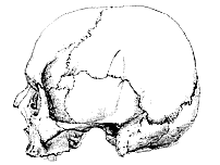
[214] The two most thoroughly contrasted human skulls, taking them altogether, which have hitherto fallen under my notice, are those various aspects and sections of which, reduced to one-third of the size of nature, are represented in the accompanying woodcuts.
The one of these skulls, which I shall call A, belongs to the Museum of the Royal College of Surgeons of England, and is thus described in the "Osteological Catalogue."
"5484. The cranium of a native of Tartary.
"It is remarkable for its breadth and shortness, slightly convex superior surface, and broad, high, and vertical occipital surface. The forehead is broad but low. The nasal bones are large and prominent; the malars are not prominent. The anterior alveoli of the upper jaw slope forwards–Hunterian."
For the opportunity of examining and making the requisite section of the other skull, B, I am indebted to J. B. Sedgwick, Esq., into whose possession it came, many years ago, as a "New Zealand" skull.
Among a good many New Zealand skulls which I have examined, however, none resemble this; while it presents so many Australian characters, that I am disposed to think it must have been obtained either in Australia, or in one of the Negrito islands. For it is a circumstance worthy of much attention, that the crania of the more or less woolly-haired Negrito inhabitants of Tasmania, New Caledonia, the Feejees, the New Hebrides, &c., present strongly Australian features, and are frequently altogether indistinguishable, by their external characters, from those of the leiotrichous Australians.
However, the precise origin of these skulls is not a matter of any moment in relation to my present purpose, which is simply to indicate [215] the nature, the extent, and the relations of the more important anatomical differences between the two skulls; and incidentally, to illustrate the mode of comparing skulls which seems to me best calculated to render their real differences apparent.
Fig. 1. The skull A. Norma lateralis .
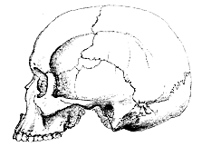
Fig. 2. The skull B. Norma lateralis .
The skull A is the broadest undistorted cranium which has come under my notice, its index being .977. It is, therefore, eminentlybrachistocephalic.1 When held out, at arm's length, so as to present [216] the norma verticalis to the eye (fig. 5), its bulging sides completely hide the zygomatic arches. It is, therefore, in Mr. Busk's nomenclature, cryptozygous.
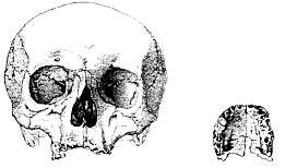
Fig. 3. The skull A. Norma facialis. Fig. 3a.
Fig. 3a. The palate of the skull A.
The squama occipitis has a well marked convexity, separated in the middle line by a depression (fig. 1) from the parietal region. A slighter depression occurs on the middle of the coronal suture; but, neither in this region, nor elsewhere, can I discover any indications of artificial distortion, unless a certain amount of asymmetry of the occiput (fig. 5), produced by the flattening of the right side (probably from nursing) can be regarded as such. The coronal suture is open, throughout its whole extent, on both the inner and the outer faces of the skull, as are the lambdoidal, occipito-mastoid, alisphenoidal, [217] squamosal, nasal and fronto-nasal sutures. But the sagittal suture is so absolutely obliterated, that not a trace of it is discernible on either the outer, or the inner, surfaces of the skull (figs. 5 and 7).

Fig. 4. The skull B. Norma faciali. Fig. 4a.
Fig. 4a. The palate of the skull B.
The squamosal and the frontal are separated on both sides, partly by the parietal and partly by an intercalary bone, which is small on the right side, larger on the left, and lies between the parietal and the alisphenoid (fig. 1).
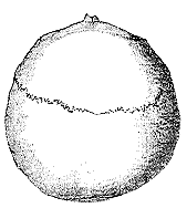
Fig. 5. The skull A. Norma verticalis .
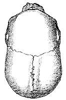
Fig. 6. The skull B. Norma verticalis.
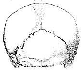
Fig. 7. The skull A. Norma occipitalis.
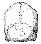
Fig. 8. The skull B. Norma occipitalis.
The auditory foramina are rounder than usual; the mastoid processes are well developed and prominent; the upper edge of the zygoma is nearly straight. The face is orthognathous, on the whole, though there is a certain amount of alveolar prognathism.
[218] Turning to the base of the skull, the obliquity of the axes of the glenoid cavities is very remarkable. If these axes were prolonged inwards, they would cut one another as far back as the junction of the middle and anterior thirds of the occipital foramen. The axis of this foramen is directed downwards and forwards.
Traces of the maxillo-premaxillary suture are visible upon each side of the naso-palatine foramen. The palato-maxillary suture is unclosed, and convex forwards in the middle line (fig. 3a).
The frontal sinuses are extensive, and are separated by an imperforate septum which lies a little to the right of the middle line. The body of the sphenoid and the roots of its lesser and greater alæ are occupied by the sphenoidal air-cells, the right cell extending back, beneath the sella turcica, to the posterior limit of the body of the basi-sphenoid.
The right second bicuspid and first true molar are the only teeth which remain, and their crowns are ground down to flat surfaces by wear. From this and other circumstances there can be no doubt that the possessor of this skull had attained middle age.
While A is the widest, B is the narrowest normal skull I have met with, its index being only .629. It is, therefore, an extremely marked example of mecistocephaly. It is, further, phænozygous, the norma verticalis exhibiting a space between the zygomatic arches and the sides of the brain case (fig. 6).
The sagittal, coronal, lambdoidal, occipito-mastoid, alisphenoidal, squamosal, nasal, and fronto-nasal sutures, are open throughout their whole extent.
The frontal and the squamosal bones remain separate on both sides. On the left side, there is a considerable intercalary bone between the alisphenoid, parietal, frontal, and squamosal (fig. 2).
The auditory foramina are vertically elongated, perhaps more so than is usual: and they are, both absolutely and relatively, narrower than those of A. The mastoid processes are well developed and prominent. The upper edge of the zygoma is slightly convex upwards.
The face is obviously prognathous.
On the base of the cranium the axes of the glenoid cavities, if prolonged inwards, would cut one another about half an inch in front of the anterior edge of the occipital foramen, so that they are nearly transverse to the long axis of the skull. The axis of the occipital foramen is directed downwards and forwards. Faint indications of the maxillo-premaxillary sutures are discernible upon both [219] sides. The maxillo-palatine suture persists, and is arched forwards in the middle line (fig. 4a). The frontal sinuses are divided by a septum, which is inclined to the left side above. They are not quite so large as in A, but they are much larger than is usual in true Australian skulls.
The large sphenoidal air-cells are divided in the middle line by a septum. They do not extend back further than the middle of the pituitary fossa. The last molar is cut and in use; but the other teeth are only slightly worn. The skull may have belonged to a person of between twenty-five and thirty years of age.
In the norma lateralis (figs. 1 and 2), beside the points already mentioned, the great difference in the longitudinal contours of the two skulls is obvious. Further, the temporal ridge, sharply marked in B, is almost obsolete in A. The supra-auditory ridge, on the other hand, is more distinct in A than in B. The occipital spine is not very strong in either skull; and in both, the contour of the squama occipitis projects beyond it when the skull is horizontal. This projection is greater in B. In the norma occipitalis (figs. 7 and 8) there is a wonderful contrast between the spheroidal dome of A and the sharply ridged, wall-sided, pentagon of B; an opposition quite as marked in its way as that between the normæ verticales (figs. 5 and 6) of the two crania. The denticulations of the sutures are, for the most part, more simple in B than in A.
Each of the skulls A and B has been longitudinally and vertically bisected, and the outlines of each section having been accurately marked upon sheets of tracing paper, the one outline has been superimposed upon the other, in such a manner, that the basicranial axes2 correspond in direction, while their anterior ends, situated at the point of junction of the presphenoid and ethmoid, coincide.
The resulting figure, reduced to one-half the size of nature, is given in fig. 9. In this figure a, b represents the basicranial axis; bc, bc lines drawn from the occipital end of the basicranial axis to the opposite boundary of the occipital foramen. The angle a, b, c, therefore, indicates the inclination of the plane of the occipital foramen to the basicranial axis, and will be small, or great, in proportion to the extent to which the occipital foramen looks forwards and downwards. It is analogous to, though not identical with, Daubenton's angle. ad, ad are lines drawn from the ethmoidal end of the basi[220]cranial axis to the anterior edge of the premaxilla, where it bounds the nasal aperture. The angle b, a, d, may be termed the premaxillary angle.
ae, ae mark lines drawn from the ethmoidal end of the basicranial axis to the middle of the posterior margins of the palatine plates of the palatine bones. The angle b, a, e, may be termed the post-palatine angle.
The line ag is drawn from the anterior end of the basicranial axis, through the upper ends of the ethmo-frontal sutures, f, f ; af, af consequently indicate the lengths of the respective cribriform plates; while ag defines the general planes of those plates; which happen in the present case, to coincide.
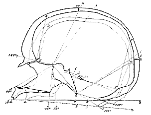
Fig. 9. Diagram exhibiting longitudinal and vertical sections of the skulls A, B, reduced to one-half the size of nature and superimposed. The thick contour lines and letters belong to B, the thin ones and the dotted lines to A.
b, a, g, is the basi-ethmoidal angle, which diminishes in proportion as the line ag rotates downwards upon a ; or, in other words, in proportion to the departure of the human skull from the condition of that of the lower Mammalia.
fk, fk are perpendiculars to the line ag erected upon the point j. [221] The distance between these lines and the inner contour of the frontal bones is a measure of the anterior cerebral overlap; or, of the extent to which the frontal lobes of the brain project beyond the extremities of the olfactory nerves. ah, ah; ai, ai, are lines drawn from the anterior end of the basicranial axis to the middle of the coronal and lambdoidal sutures Cr, L.
lC, lC are lines drawn from l, l, the points at which the inner end of the posterior superior margin of the petrosal cuts the basicranial axis, to the torcular Herophili. Each may be taken to represent the tentorial plane, though, of course, the centre of the tentorium would rise considerably above it. Cv is a perpendicular erected upon the hinder end of this line and marking the projection of the cerebral hemispheres beyond the cerebellum, or the posterior cerebral overlap, AB is a line representing the extreme length of the entire cranium; y, its centre; x, the point cut by a perpendicular from the centre of the occipital foramen, Au the internal auditory foramen, L the lambdoidal, Cr the coronal suture.
The various measurements of the two skulls, to which I shall have to advert, may be conveniently classified under three heads: 1stly, those which are identical; 2ndly, those which differ by not more than five per cent.; 3rdly, those which differ more than five per cent.
They are given in 1/100ths of an inch, not from any desire to affect an accuracy to which cranial measurements can lay no claim, but for the sake of more ready comparison. It must be understood that very few cranial measurements will come out exactly alike twice over; so that the numbers given must in most cases be regarded simply as a mean; the variations from which will not affect the general result.
| I. Identical measurements (in 1/100ths of an inch). | ||||
| A. | B. | |||
| 1. The basicranial axis | 235 | 235 | ||
| 2. The vertical height of the face from the fronto-nasal suture to the alveolar margin | 260 | 260 | ||
| 3. The vertical height of the orbital aperture | 135 | 135 | ||
| 4. Anterior interlachrymal diameter, from the point of junction of the frontal, lachrymal and maxillary, on the one side, to that on the other | 75 | 75 | ||
| 5. From the margin of the orbit to the alveolar margin between m1 and m2 | 175 | 175 | ||
| 6. The greatest breadth of the palate taken between the inner edges of the alveoli | 140 | 140 | ||
| 7. The greatest length of the palatine plate of the palate-bone | 75 | 75 | ||
| [222] II. Measurements which do not differ more than 5%.
Differences in favour of B are marked with a *. | ||||
| A. | B. | Diff. | ||
| 8. The longitudinal arc of the frontal | 512 | 537 | *25 | |
| 9. The longitudinal arc of the occipital | 430 | 426 | 4 | |
| 10. The greatest transverse diameter of the occipital bone from one occipito-mastoid suture to the other | 443 | 422 | 21 | |
| 11. The length of the occipital foramen | 145 | 140 | 5 | |
| 12. The distance of the suborbital foramina | 230 | 220 | 10 | |
| 13. The length of the zygoma from the anterior edge of the auditory foramen to the anterior end of the maxillo-jugal suture | 310 | 323 | *13 | |
| III. Measurements which differ more than 5%. | ||||
| 14. Extreme length | 670 | 755 | *85 | |
| 15. Extreme breadth | 6553 | 4754 | 180 | |
| 16. Height | 480 | 530 | *50 | |
| 17. Longitudinal arc of the parietals | 450 | 550 | *100 | |
| 18. Transverse arc from one auditory foramen to the other | 1350 | 1175 | 175 | |
| 19. Width of the frontals immediately behind the external orbital process (least frontal measurement) | 405 | 340 | 65 | |
| 20. Width of the frontals on the temporal ridge just above the external orbital process | 417 | 375 | 42 | |
| 21. The greatest frontal width, where the temporal ridge cuts the coronal suture | 555 | 395 | 160 | |
| 22. Length of the cribriform plate | 95 | 107 | *12 | |
| 23. Posterior interlachrymal diameter, between the junctions of the ethmoid, lachrymal and frontal .. | 105 | 90 | 15 | |
| 24. Between the posterior ends of the ethmo-maxillary sutures | 170 | 155 | 15 | |
| 25. Between the outer edges of the optic foramina in the interior of the skull | 126 | 85 | 41 | |
| 26. Between the outer edges of the optic foramina in the orbits | 155 | 115 | 40 | |
| 27. Between the outer sides and posterior edges of the bases of the external pterygoid processes | 205 | 176 | 29 | |
| 28. Between the points of the alispheno-squamosal sutures, which are cut by the transverse ridge on the alisphenoid | 360 | 320 | 40 | |
| 29. Between the outer edges of the foramina ovalia | 240 | 193 | 47 | |
| 30. Between the posterior ends of the alispheno-squamosal sutures and outer sides of spinous processes | 305 | 245 | 60 | |
| 31. Between the outer edges of the glenoidal fossæ | 533 | 455 | 78 | |
| 32. Between the most distant points of the outer surfaces of the mastoid processes | 536 | 452 | 84 | |
| 33. Transverse arc of the occipital, from the junction of the lambdoidal suture and its additamentum on one side, horizontally over the occiput to the other side | 500 | 636 | *136 | |
| 34. Between the centres of the styloid foramina | 360 | 290 | 70 | |
| 35. Least breadth of the basicranial axis (between the apices of the partes petrosæ) | 100 | 75 | 25 | |
| [223] 36. Between the inner edges of the precondyloid foramina | 150 | 125 | 25 | |
| 37. Between the most distant points of the outer edges of the occipital condyles | 215 | 185 | 30 | |
| 38. Transverse diameter of the occipital foramen | 125 | 105 | 20 | |
| 39. Between the internal auditory meatuses | 245 | 155 | 90 | |
| 40. Length of the posterior superior edges of the ossa petrosa | 270 | 245 | 25 | |
| 41. Extreme transverse distance of the outer surfaces of the zygomata | 555 | 485 | 70 | |
| 42. From the lower end of the basicranial axis to the anterior alveolar margin of the pre-maxilla | 332 | 420 | *88 | |
| 43. From the lower end of the basicranial axis to the posterior end of the spine of the palate | 137 | 165 | *28 | |
| 44. Extreme distance of the outer surfaces of the maxillæ | 220 | 242 | *22 | |
| 45. Width of the external nasal aperture | 92 | 105 | *13 | |
| 46. Greatest perpendicular height of the nasal passage, from the cribriform plate to the upper surface of the palate | 177 | 160 | 17 | |
| 47. Extreme distance of the outer walls of the posterior nares | 115 | 98 | 17 | |
| 48. Height of the arch of the posterior nares | 100 | 92 | 8 | |
| 49. From the posterior end of the spine of the palate to the anterior margin of the pre-maxillæ | 195 | 253 | *58 | |
| 50. Length of a line drawn from the anterior edge of the premaxilla along the posterior edge of the occipital foramen, and cut by a perpendicular tangent to the occiput, A, B. fig. 9 | 660 | 810 | *150 | |
| 51. Length of so much of this line as lies in front of a point cut by a perpendicular to the centre of the occipital foramen (A, x) | 390 | 480 | *90 | |
| 52. Length of the line behind this point (x, B) | 270 | 330 | *60 | |
| 53. Projection of the inner contour of the frontal beyond the level of a perpendicular to the anterior end of the cribriform plate (= anterior cerebral overlap) | 20 | 55 | *35 | |
| 54. Projection of the inner contour of the occipital beyond the torcular Herophili (= posterior cerebral overlap) | 30 | 85 | *55 | |
| 55. The capacity of the cranium and volume of the brain, as determined by the volume of a cast of the interior of the skull, in cubic inches | 95 | 80 | 15 | |
| 56. Extreme length of the cast | 625 | 695 | *70 | |
| 57. Extreme breadth | 615 | 455 | 160 | |
| 58. Breadth: length = 100 | 98 | 65 | 33 | |
1. The first point to be noted in comparing the skulls A and B, the most important measurements of which have now been given, is the equality of length of their basicranial axes, which proves that brachycephaly and dolichocephaly are not necessarily connected with the shortening or the lengthening of the base of the skull; but that their most extreme forms may arise exclusively from modification of the side walls and roof of the cranium. In the present case, the difference in the absolute lengths of the two skulls amounts to about 11% of the length of the longer (B). It is due, in part, to the remarkable shortness of the parietal region in A, the longitudinal arc of the parietals being about 18% (No. 17), and its chord 20%, longer in B [224] than in A. In part, the superior length of B arises from the forward extension of the frontal region, which will be more particularly discussed below. The elongation of the occipital region does not seem to be greater than that which would necessarily accompany the lengthening of the parietals.
The difference in breadth between A and B (No. 15) amounts to 27% of the broader. It is greater absolutely and relatively than the sum of the excess of height and length of B over A.
2. Virchow's "sattel-winkel" is substantially the same in the two skulls, though the one is vastly more prognathous than the other. Hence it follows that there is no necessary connection between the "sattel-winkel" and prognathism or orthognathism.
3. It will be observed, that only one pair of the transverse measurements, which differ less than 5%, appertain to the brain-case. These are No. 10, the greatest transverse diameters of the occipital bone. In all the other transverse measurements of the brain-case (with the exception of No. 33, the transverse arc of the occipital, which is in reality as much a longitudinal as a transverse dimension) A exceeds B. Hence it appears that, in such a thorough example of brachycephaly as this, the excess of transverse growth is general, and affects all parts of the brain-case. But the excess is not equal in all regions of the skull.
| Thus in No. | 35 | the excess of A is about | 25% |
| ... | 39 | ... | 36% |
| ... | 36 | ... | 16% |
| ... | 34 | ... | 19% |
| ... | 32 | ... | 15% |
| ... | 31 | ... | 14% |
| ... | 30 | ... | 20% |
| ... | 29 | ... | 19% |
| ... | 28 | ... | 11% |
| ... | 27 | ... | 14% |
| ... | 25 | ... | 33% |
| ... | 21 | ... | 29% |
| ... | 19 | ... | 16% |
| ... | 15 | ... | 27% |
The measurements which exhibit the smallest difference (No. 28), represent roughly the distances of the apices of the temporal lobes of the brain. The greatest differences are shown by the base of the brain in the region of the pons and anterior part of the medulla (No. 39); by the exits of the optic nerves (No. 25); by the region corresponding with the outer sides of the frontal lobes (No. 21); by the region corresponding with the outer sides of the parietal lobes, [225] or the posterior and upper parts of the temporal lobes (No. 15); and by the width of the basicranial axis itself.
It will be very interesting to ascertain, from similar measurements of other skulls, to what extent the rule observed in these, that in skulls with equal basicranial axes5 dolichocephali are absolutely narrower than brachycephali in their transverse diameters, holds good. Even in the present cases there is the remarkable exception with regard to the transverse diameters of the occipital (No. 10), which has already been noted; and it is, of course, quite conceivable that the diameters of the base of the cranium should vary irrespectively of those of its side walls. But a skull which should derive its excess in breadth from a development of its side walls alone, exhibiting what might be called lateral brachycephaly, would obviously have a different significance from one which, like A, is basally, as well as laterally, brachycephalic. And similar considerations apply to dolichocephaly.
4. By "vertical height" in the foregoing table of measurements, I mean the distance from the posterior and inferior end of the basicranial axis to the point of intersection of the corona and sagittal sutures. These are convenient fixed points, and although a line joining them is by no means constantly perpendicular to a longitudinal axis of the skull, or inclined at any invariable angle to the basicranial axis, yet, practically, it hardly ever differs more than a tenth of an inch from a more strictly vertical measurement.
The vertical height of B exceeds that of A by about 10%; but while A is within 0.1 inch as low as any skull I have met with, B is not more than half way towards the maximum of height which I have observed, and which is about an inch greater than that of B.
The following table of a few measurements of sections of crania seems to show that the height of skulls as thus estimated, varies without much reference to their other measurements.
| Length of the basicranial axis. | Height. | Cephalic index. | |
| 1. Scutari (Turk? 5563A) | 245 | 575* | .88 |
| 2. Japanese | 290 | 570 | .75 |
| 3. Redondo (Negro) | 275 | 570* | .72 |
| *** The * indicates that the height of the parietal region exceeds that given by about 0.1 inch. The remarkable length of the basicranial axis in Nos. 2, 3, and 7 must be taken into account. The numbers refer to the Catalogue of the Museum of the Royal College of Surgeons. | |||
| [225] 4. Australian (5317) | 250 | 565 | .68 |
| 5. Tasmanian (5324) | 225 | 550 | .76 |
| 6. English (5733) | 250 | 550 | .74 |
| 7. Japanese | 272 | 550* | .75 |
| 8. Australian (5307) | 250 | 545 | .69 |
| 9. The skull B | 235 | 530 | .629 |
| 10. Australian | 230 | 530 | |
| 11. Chinese (5474) | 235 | 535* | .75 |
| 12. Malay (5463A) | 240 | 510* | .88 |
| 13. Japanese | 240 | 490* | .76 |
| 14. Australian (5331) | ? | 490 | .71 |
| 15. Mondombe (Negro) | 230 | 480 | .74 |
| 16. The skull A | 235 | 480 | .977 |
5. Certain important differences between the skulls A and B, which would not be brought out by the ordinary methods of measurement and comparison, become very obvious in the sectional diagram (fig. 9). Thus, the spaces behind the lines Cv, Cv, and in front of f, k, f, k are very much greater in B than in A, showing that the cerebral hemispheres overlapped the cerebellum behind and the cerebrum in front, for a much greater distance in B than in A. The distance between the anterior contour of the cranial cavity and the anterior end of the basicranial axis in B is further increased by the greater length of the cribriform plate a, f . Hence, if we call the space comprised between the lines a, h, a, f, and the anterior inner contour of the frontal bone, the frontal area of the longitudinal section, this frontal area is absolutely greater and projects further forwards in B than in A. The difference between the two skulls thus produced is further exaggerated by a sort of rotation of the whole cranial chamber upon its axis, forwards in B and backwards in A. Thus the whole forehead, with the coronal suture, is thrown backwards in A, and the occipital plane, partaking in the same movement, forms a more open angle with the basicranial axis than that of B. The tentorial plane has shifted in the same sense to a less degree. The line b, i, in A, on the other hand, is a little in advance of the corresponding line in B, probably in consequence of the remarkable brevity of the parietal bones.
The skulls A and B present as complete a contrast as any I have seen in regard to this remarkable rotation of the skull upon its basal axis. But it must not be supposed that the backward rotation is connected with brachycephaly, or the forward rotation with dolichocephaly. I have sections of two dolichocephalic Australian skulls [227] which differ as widely as A and B in the anterior region of the skull. I have other sections of brachycephalic skulls, in which the frontal contour lies far in advance of that of A, though I have not yet met with a brachycephalic skull having so great a forward rotation of the frontal region as B.
As a rule, the coronal suture is situated forward with forward rotation of the skull; backward, in the contrary case. But neither the lambdoidal suture, nor the posterior edge of the occipital foramen, necessarily follows it. In a negro skull, with nearly the same extent of backward rotation of the frontal region as in A, the line b c makes an angle of only 135° with a b, or 26° less than that in the case of A.
The influence of the backward and forward rotation of the frontal region of the skull upon orthognathism and prognathism, as they are ordinarily estimated, is obvious. The so-called facial angle, in fact, does not simply express the development of the jaws in relation to the face, but is the product of two factors, a facial and a cranial, which vary independently. The face remaining the same, prognathism may be indefinitely increased, or diminished, by the rotation of the frontal region of the skull, backwards or forwards, upon the anterior end of the basicranial axis.
If B had the frontal contour of A, it would be an extremely marked example of orthognathism, or rather of what Weicker has called "opisthognathism"; while, if A had the frontal contour of B, it would appear to be marvellously prognathous. And yet in neither case would there be any change in the jaws, but only so much modification in the position of the cranial cavity relatively to its axis, as can be shown to occur among skulls belonging to the same stock.
6. The real differences in the disposition of the facial bones relatively to the basicranial axis, or, in other words, the amount of true prognathism or orthognathism, cannot, in fact, be safely estimated by any of the accepted "facial angles." The sectional diagram shows that B is truly much more prognathous than A, the differences between the two being of three kinds.
(a) The vertical height of the nasal cavity is somewhat less in B than in A.
(b) The length of the palate is greater in B than in A.
(c) Lines drawn from the anterior end of the basicranial axis to the posterior and anterior margins of the floor of the nasal cavities (a e, a d) form more open angles (premaxillary and postpalatine angles) with a, b in B, than they do in A. In other words, the centre of the palate has, so to speak, moved forwards in B.
Increase in the absolute length of the palate and shifting forward [228] of the centre of the palate, are competent, singly or together, to produce true prognathism, other conditions remaining unaltered. But shortening of the vertical height of the nasal chamber alone is consistent with the preservation of complete orthognathism. As a general rule, however, the three conditions are concomitant, as in A and B ; the more prognathous skull having a lower nasal chamber as well as a longer palate, and a shifting forward of the centre of the palate.
Practically, I should say that the angle b, a, d fairly represents the degree of true prognathism; and I think it will be found convenient to consider skulls in which that angle is less than 95° as orthognathous, and those in which it is greater as prognathous. The most prognathous skull I have met with had the angle b,a, d = 110°; in the most orthognathous it was only 83°. I doubt if the angle ranges much beyond 30°.
7. The vertical measurements of the face (Nos. 2, 3, 5, 46, 48) either agree, or differ but little, the excess being in favour of A (Nos. 46, 48). Among the longitudinal measurements, those of the palatine plates of the palatine bones agree (No. 7), showing that the difference in the lengths of the palates is wholly due to the premaxillæ and maxillæ. Again, the lengths of the zygomata differ only to an inconsiderable extent, whence it follows that the excess of longitudinal growth is in the alveolar, rather than in the orbital parts of the maxillæ. No. 42 shows that the margin of the premaxilla is .88 inch more distant from the lower end of the basicranial axis in B than in A ; but No. 43 proves that 0.28 of this amount is due, not to the excessive growth of the maxilla and premaxilla, but to the shifting forward of the palate as a whole.
8. Taking the length of a line A, B, drawn from the anterior alveolar margin of the premaxilla along the posterior edge of the occipital foramen and cut by a perpendicular tangent to the posterior face of the occiput, to represent the basal length of the whole skull (No. 50), it is much longer (1.5 inch) in B than in A . Nevertheless, the extra length of B is so distributed that the centre of the occipital foramen (x) is in exactly the same place in the two skulls, viz. at three-fifths of the length of the line A, B from its anterior end.
9. Although A is cryptozygous and B phænozygous, the inter-zygomatic width of the face is greater in A than in B by about 12% (No. 41). But the maxillary diameter (No. 44), on the contrary, is about 9% greater in B than in A. This arises, however, chiefly from [229] the less development of the alveoli themselves in A, seeing that the breadth of the palate inside the alveoli (No. 7) is the same in the two. The distances of the suborbital foramina (No. 12) are nearly the same in the two skulls, and those of the upper ends of the ascending processes of the maxillary are identical. Hence the greater width of the face in A is not due to any excess in the size of the bodies of the maxillary or premaxillary bones, but to the increased transverse diameter of the frontals (Nos. 19, 20), which push the jugal bones outwards. This is accompanied by a certain increase in the diameter of the ethmoid (Nos. 23, 24), of the distance between the optic foramina (No. 26), and of the width of the posterior nares (No. 47).
10. In describing the general characters of the two skulls I have mentioned the interesting fact that the chief sutures of B, the very long skull, are completely open; but that the sagittal suture of A, the very short and broad skull, is thoroughly obliterated, while the other great sutures named are still open.
It is therefore clear that extreme brachycephaly is consistent with comparatively early synostosis of the parietal bones; or, in other words, that synostosis of those bones may take place comparatively early and yet have no discernible effect upon the form of the skull.
This is, in fact, perfectly obvious from the nature of the case. For the final proportions of the brain-case of the human skull are attained in early manhood, while the sagittal suture ordinarily remains open till late in life; and it can make no manner of difference to the shape of the brain-case whether the sagittal suture becomes obliterated at thirty, or at fifty, years of age, if the braincase assumes its final proportions at twenty-five.
When the skull of a man of middle age, of unknown stock, with obliterated sagittal suture, is placed before an anatomist, he possesses absolutely no evidence respecting the period at which the obliteration took place; and, consequently, he has no means of judging whether the synostosis has, or has not, had any share in producing the form of the skull. If the cephalic index of the skull be greater than .70, he has not the least right to suppose that the synostosis has had any effect, inasmuch as there is abundant evidence to prove that crania with lower cephalic indices exhibit no such synostosis.
The Neanderthal skull belongs to a man of middle age, is of unknown stock, and has a cephalic index of .72. There is consequently not a shadow of justification for the assumption that any [230] obliteration of the sagittal suture which it presents has had more effect in narrowing its proportions, than the obliteration of the sagittal suture in A has had upon the configuration of that exceedingly broad skull.6
See "Notes upon the Human Remains from Keiss," in Mr. Laing's Prehistoric Remains of Caithness, p. 85. 2 By basicranial axis, I mean a line drawn through the middle vertical plane of the basi-occipital,
basisphenoid and presphenoid, from the hinder extremity of the former bone to the anterior extremity of the last, at the upper end of the ethmo-presphenoid suture. 3 Between the upper edges of the squamosals, over the auditory foramina. 4 Between the parietal tuberosities. 5 In comparing skulls the length of the respective basicranial axes must be carefully taken into account. Had the basicranial axes of A and B been unequal I should have given all the measurements of each skull in terms of its own basicranial axis; but as they are equal, I have not thought it worth while to take the trouble of making the requisite calculations. 6 With the permission of the Museum Committee of the Royal College of Surgeons, and of Mr. Sedgwick, casts of the skulls A and B and of their cavities, representing the corresponding brains, have been made and are to be obtained of Mr Gregory, Russell Street, Covent Garden.
1 Cephalic index at or above .80 = Brachycephali, round skulls. .85 = Brachistocephali. below .85 and .80 = Euryocephali. .80 = Dolichocephali . .80 .77 = Sub-brachycephali }oval skulls. .77 .74 = Orthocephali. .74 .71 = Mecocephali. .71 = Mecistocephali, oblong skulls.
|
THE
HUXLEY
FILE
|
| ||||||||||||||||||||||||||||||||||||||||||||||||||||||