[493] The brain of a Spider Monkey (Ateles belzebuth) has already been partially described and figured by M. Gratiolet in his remarkable memoir 'Sur les Plis Cérébraux des Primatès' (1854); but this careful observer had only old spirit specimens at his disposal, and it did not enter into his plan to give any account, either of the internal structure of the cerebrum, or of its relations to the cerebellum, or of the cerebellum itself. Hence a new description, which should touch upon these points, could hardly be superfluous, under any circumstances; while, at the present moment, the controversy which has arisen respecting the nature and the extent of the differences in cerebral structure between Man and the Apes gives an especial value to all new facts.
It has been affirmed–and a proposed new classification of the Mammalia has been largely based upon the assertion–that the brain of Man is distinguished from that of all Apes by possessing a posterior lobe, a posterior cornu to the lateral ventricle, and a hippocampus minor–these structures being absent in all Apes, even the highest.1
I have elsewhere2 exposed the fallacy of these distinctions as applied to the Apes in general; Dr. A. T. Thomson3 and Dr. Rolleston4 [494] have proved the existence of the three structures referred to in the Chimpanzee and the Orang, by investigations upon the brains of these animals, undertaken with especial reference to the questions under discussion; and I propose to continue the process of rectification thus commenced, by inquiring into another special case–that of Ateles paniscus–and proving, by direct demonstration of the facts, that the three structures, said to be absent even in the highest Apes, are, on the contrary, largely developed5 in this comparatively low American monkey, possessed of but a rudimental thumb upon its hand, and provided with four more teeth than the Old World Apes and Man.
In fact, so far from its being true that the differences between Man and the Apes lie mainly in the cerebral characters, so often referred to, all the evidence now accumulated tends towards the belief that the only three, very striking, cerebral characters, absent in other Mammalia, which can be truly affirmed to be common to Man and the Old and New World Simiæ, are exactly these three,–the whole of the true Apes, so far as our present knowledge goes, possessing a posterior lobe, a posterior cornu to the lateral ventricle, and a hippocampus minor in that posterior cornu; while these structures, so far from being in a rudimentary condition, are often more largely developed, in proportion to other parts of the brain, in the Apes than in Man.
The figures 1 and 2 of Plate XXIX represent the brains of a male and of a female Ateles of about the same size, as seen from above: both figures were drawn under my own eye by a very competent artist, and are in all essential respects perfectly faithful. It is nevertheless obvious that they differ greatly–so much, in fact, that they might readily be supposed to have belonged to different species. The whole difference, however, is due to the circumstance that, while fig. 1 was drawn from an almost fresh brain, fig. 2 represents a brain which has been for several months in spirit.6 The roundness of outline of the latter as compared with the former, [495] and the more transverse direction of the fissure of Rolando, are very remarkable; for the skulls of the two specimens show no particular difference of form. In the unaltered brain, figs. 1, 3, 4, the narrowness of the frontal lobes anteriorly, the excavation of their orbital faces, and the flatness of the superior contour are especially worthy of notice. Viewed from above, no part whatsoever of the cerebellum is visible, either at the sides or behind; while a profile view shows that the cerebral hemispheres projected, for at least 1/16th of an inch, behind the posterior edge of the cerebellum. Whether this represents the total amount of cerebral overlap or not, I cannot say, in the absence of a vertical section of an Ateles' skull; but it is amply sufficient to prove that, even accepting as the definition of the posterior lobe the novel formula "All that part of the hemisphere which covers the posterior third of the cerebellum and passes behind it," Ateles is provided with a well-developed posterior lobe.
In this respect, as I have already said, it resembles all the Old and New World Simiæ, which have yet been examined,–the only genus, within my knowledge, which even comes near to presenting an exception being Mycetes. I have not, indeed, had the opportunity of dissecting the brain of this monkey (nor has M. Gratiolet been enabled to give any account of it); but the Curator of the Hunterian Museum having kindly permitted me to have a vertical longitudinal section of the skull of a Mycetes made [sic], I found not only that the plane of the tentorium (and consequently the inferior margin of the posterior lobes of the cerebrum) had a much greater inclination than in any other Simian (making an angle of as much as 45° with the base of the skull), but that the cerebral overlap, measured in the manner described by me in the Athenæum for April l3th, 1861, does not exceed 1/20th of an inch, though the maximum length of the cranial cavity is 2.4 inches. Notwithstanding this reduction of the posterior lobe, however, the contrast between Mycetes, as a true Simian, and a Lemur is very striking, especially if both be simultaneously compared with some lower Mammal, such as the Dog. The occipital foramen in Mycetes is situated altogether upon the posterior face of the skull, and the condyles look completely backwards, as in the Dog; while the occipital crest is placed as near the postero-superior margin of the skull as in that animal. In both, the posterior face of the skull looks backwards, and not appreciably downwards. But in the Monkey the inclination of the tentorium, large as it is, is far less than in the Dog. The inner face of the occipital bone beneath the tentorium is not excavated, and the cerebral lobes projected beyond the cerebellum when the palate was horizontal. In [496] the Dog, on the contrary, the internal surface of the occipital bone below the tentorium is much excavated; and, when the palate was horizontal, the posterior edge of the cerebellum must have projected far beyond the cerebral lobes.
In Lemur catta the inclination of the tentorial plane is hardly greater than in Mycetes ; but if the palatal line be made horizontal, it will be found that the posterior boundary of the cerebellar chamber projects for 1/5th an inch beyond that for the cerebrum, although the greatest length of the cranial cavity is only 1.9 inch. In fact, the cerebral hemispheres of the Lemur have a less backward development than those of the Dog. I believe that all the Lemurs are in the same case, and that the Prosimiæ are sharply defined from the Simiæ by the fact of always having more or less of their cerebellum uncovered; so that, by this character alone, the Lemurine brain is far more widely separated from that of any Simian, than the latter is from the human brain.
While one American Monkey (Mycetes) is, if the development of its posterior lobes only be taken into account, at the bottom of the series of Simiæ, if the same character alone be considered, another Simian, inhabiting the same geographical area, is at the top; I refer to Chrysothrix sciureus, whose posterior lobes, as I. G. St.-Hilaire long ago proved,7 are better developed than those of any other Mammal, overlapping the cerebellum by one-fifth of their length. In fact, if the Primates were arranged according to the development of their posterior cerebral lobes we should have some such descending series as the following:–Chrysothrix, Cebus, Troglodytes, Man, . . . . Mycetes–a series which sufficiently illustrates the classificatory value of these structures. So much for the posterior lobe. I turn now to the next point, the demonstration of the existence of the posterior cornu in Ateles.
When the lateral ventricle was exposed in the ordinary way (Pl. XXIX. fig. 5) a straight line passing from the extremity of the anterior to that of the posterior cornu measured 2.1 inches. A distance of 1.3 inch separated the anterior end of the anterior cornu from the commencement of the descending cornu; while a straight line extending from the commencement of the descending to the end of the posterior cornu measured 0.75. Each lateral ventricle, measured from the centre of the corpus callosum to the outer boundary, at its widest point, or opposite the commencement of the descending cornu, was about half an inch wide. The posterior cornu has a general direction backwards, outwards, and then [497] inwards; and, besides its general curvature, it has a secondary inflexion, so as to be a little sinuous. It is wide at its commencement, but rapidly narrows, until, where it bends inwards, its walls are so close together as to give it the appearance of a mere fissure, whose sides are apt to adhere together in such a manner as seriously to interfere with the satisfactory definition of the posterior limits of the cornu. In preparing the specimen, of which fig. 5 is a representation, for the artist, I therefore took care not to extend these limits artificially, rather preferring to leave a portion of the cornu unopened than to exaggerate its length.
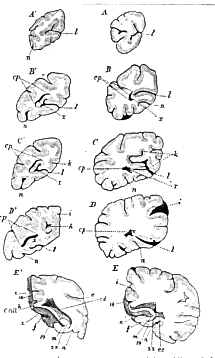
In the other brain I found the posterior cornu, on the right side (dissected in the ordinary manner), to be traceable, without the least difficulty, to within a very short distance of the posterior limit of the hemisphere; while in the left hemisphere, which I examined by making successive vertical sections from behind forwards, the posterior cornu ended at fully a quarter of an inch distance from the posterior extremity of the hemisphere. Such sections are of particular value; for they show the extent of the cornu without any disturbance of its natural dimensions; and a comparison of the woodcuts (fig 1) A, B, C, &c., and A', B', C', &c., which represent two series of sections of corresponding regions of the Human and the Ateles' brain, will at once show that the relative dimensions of the posterior cornu are greater in the Monkey than in Man. I may remark that, of the left hemispheres of three human brains which I have dissected for comparison with Ateles, that whose sections are represented in the figures had its posterior cornu far better developed than the other two, in one of which the cornu was a mere fissure, while in the other it was excessively short, not extending for more than half an inch behind the corpus callosum.
Thus, not only does the posterior cornu of the lateral ventricle exist in Ateles ; not only has it that backward, outward, and then inward curvature which has been wrongfully asserted to be peculiar to the homologous cavity in the human brain; but it is, in proportion, wider than in the human brain, and it is longer than in many human brains.
The third point in my argument is the demonstration of the existence of the hippocampus minor. But such strange confusion has been lately introduced into anatomical science, partly by a misapplication of well-understood terminology, and partly, to all appearance, by a want of proper acquaintance with the structure and nomenclature of the human brain, that I must begin ab initio, by a description of the latter, so far as regards the hippocampi and their related structures.
[498] The term "Hippocampus minor" was first used by Vicq d'Azyr in the following passage of his famous Traité d'Anatomie et de Physiologie (tome i. 1786), where, in the Explication des Planches du Cerveau, pl. 6, p. 9, I find:–"26. 46. 45. Saillie ou relief qui se continue en 26 avec l'origine de la corne d'Ammon, et qui en 45 se recourbe en dedans: c'est la partie que Morand a appellée l'ergot."8
The term "hipppocampus minor" has been used in the sense here defined by Vicq d'Azyr by all succeeding anatomists, as the following extract from the celebrated work on Human Anatomy, "Soemmering, vom Baue des menschlichen Körpers," Bd. IV. (Hirn-und Nervenlehre, umgearbeitet von G. Valentin) pp. 195, 196, will show:–"Der Sporn, oder die Klaue, oder der Vogelsporn, oder die Vogelklaue, oder der kleine Fuss des Seepferdes, oder der Nagel, oder der Stiefel, oder die Falte, oder der Hanensporn, oder die hintere, oder kleinere, Wulst, oder die fingerförmige Erhabenheit (calcar s. unguis, s. calcar avis, s. hippocampus minor, s. pes hippocampi minor, s. eminentia minor, s. digitata, s. unciformis, s. ocrea, s. colliculus), bildet eine nach aussen und vorn convex gebogene Erhabenheit der inneren Wand des hinteren Hornes des Seitenventrikels."
"The hippocampus minor forms an elevation, convex outwards and forwards, of the inner wall of the posterior cornu of the lateral ventricle."
There can, therefore, be no doubt as to what is meant by the term 'hippocampus minor.'
Another elevation of the wall of the ventricle is known to human anatomists as the 'Eminentia collateralis,' for an authoritative definition of which I will again quote Soemmering's Anatomy, "Die seitliche Erhabenheit oder die längliche Seitenerhabenheit oder die Nebenerhabenheit (eminentia lateralis, s.collateralis, s. Meckelii), bildet eine wulstige Hervorragung welche vor dem Eingange in das hintere und neben dem in das untere Horn des Seitenventrikels liegt, und nach aussen von dem Ammonshorne sich befindet. Uebrigens wird diese Benennung offenbar auf verschiedene, variabele, grossere oder unbedeutendere, Erhabenheiten, die neben dem Ammonshorne, in dem
[499]

Fig. 1.–Transverse sections of corresponding (left) cerebral hemispheres of Man (A, B, C, D, E) and of Ateles (A', B', C', D', E'), taken perpendicularly to the plane of the corpus callosum, along the lines marked with corresponding letters in Fig. 2. l. calcarine sulcus; n. collateral sulcus; i. calloso-marginal; k, occipito-parietal sulcus; c. p. posterior cornu; x. hippocampus minor; xx. hippocampus major. A-D, A'-D' viewed from behind; E and E' from in front.
[500] Bereiche des unteren Horses des Seitenventrikels vorkommen, angewendet."
"The eminentia lateralis, or collateralis, or Meckelii, is formed by a rounded elevation which lies in front of the entrance into the posterior, and beside that into the inferior cornu of the lateral ventricle, and is situated external to the cornu Ammonis."
It will be observed that Valentin, who has taken great care to collect together the multitudinous synonyms of the parts of the brain, does not enumerate "pes hippocampi minoris" among those of the eminentia collateralis; nor has the term 'pes hippocampi minoris' been ever used in this sense by any anthropotomist of authority.
And if it be an error in terminology to apply the name of pes 'hippocampi minoris' to the eminentia collateralis, it is a still greater error, in point of anatomical fact, to assert that "the eminence continued backwards from the pes into the posterior cornu is the hippocampus minor."9 If any eminence is continued backwards from the eminentia collateralis into the posterior cornu (as sometimes happens) it lies in the floor of the cornu, alongside the hippocampus minor, but perfectly distinct from it. But it will perhaps be better to demonstrate this elementary fact over again, though I feel that the doing so necessitates an apology to those who are conversant with the anatomy of the human brain.10
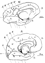
The lower figure of the woodcut (fig. 2) represents the inner surface of one of the hemispheres of the human brain. The contour is taken from one of Foville's Plates, but only the principal sulci are indicated,–those marked l, m, and n being put in from a specimen which I dissected, so as to ascertain their true nature. Of these sulci, that marked i i is the sulcus called by Gratiolet 'fronto-parietal,' a name which involves an ambiguity, and for which I therefore propose to substitute 'calloso-marginal,' as this sulcus lies between the corpus callosum and the margin of the hemisphere; k is the occipito-parietal sulcus (scissure perpendiculaire interne, Gratiolet); l is the posterior part of the "scissure des hippocampes" of Gratiolet. This sulcus is a very remarkable one. Commencing just in front of the posterior thickening of the corpus callosum, opposite x, it rapidly deepens as it [501] is traced backwards, and forms a great fissure, extending, in some parts, for as much as 3/4 of an inch upwards and outwards, and passing backwards until it nearly reaches the posterior margin of the hemisphere, where it terminates by dividing into two short, but deep, branches, a superior and an inferior. Traced from before backwards, or from within outwards, the line of this sulcus presents a strongly marked, but irregular, upward convexity.

Fig. 2.–View of the inner surface of the left cerebral hemisphere of Ateles, of the natural size, and beneath it a corresponding view of the human left cerebral hemisphere reduced to the same size. In the latter only the principal sulci are indicated. i. calloso-marginal sulcus; k. occipito-parietal sulcus; l. calcarine sulcus; m. dentate sulcus; n. collateral sulcus; x. continuation of the callosal gyrus (18) into the uncinate gyrus (19). A, B, C, &c., A', B', C', & c., the lines along which the transverse sections in Fig. 1 are taken.
On making successive transverse sections of this cerebrum from before backwards (woodcut, fig. 1. A, B, C, D), the fissure was seen, in its most posterior part (A), to pass almost horizontally outwards for a short distance, and then to divide into an upward and a downward branch. In front of A it forms a curve strongly convex upwards, without any terminal bifurcation; in B it is much longer and less convex; in C it is but slightly sinuous, and in D it is a little concave [502] upwards and inwards. Combining these views with those given in fig. 2, it is easy to form an estimate of the figure of the surfaces of the upper and under lips of the sulcus; but what is most important about it is, that, so far as the posterior cornu extends, the closed end of this sulcus corresponds with the hippocampus minor (x), which last is, in truth, nothing but the arch of cerebral substance which, at once, forms the outer boundary of the sulcus and the inner boundary of the cornu.
From its special relation to the hippocampus minor, or "calcar avis," I shall call this the "calcarine" sulcus; but it extends beyond the calcar and the posterior cornu, both anteriorly and posteriorly, particularly in the latter direction. Nevertheless it does, in a definite sense, correspond with the inner wall of the posterior cornu. The calcarine sulcus dies away anteriorly, at the point indicated, and is in no way continuous with that sulcus which has a relation to the hippocampus major similar to that of the calcarine sulcus to the hippocampus minor, and which, for distinction's sake, I will call the 'dentate' sulcus, on account of its relation to the fascia dentata or corps godronné . This narrow and well-known sulcus lies between the letters m and m, the lower m being placed opposite its termination in the fold formed by the recurved part (crochet de l'hippocampe, Gratiolet) of the so-called 'uncinate' convolution (19). Thus the dentate sulcus, which corresponds with the hippocampus major, is separated from the calcarine sulcus, which similarly answers to the hippocampus minor, by the rounded process of cerebral matter, x, this last being, in fact, the inferior and posterior continuation of the callosal gyrus (circonvolution de l'ourlet of Foville, pli du corps calleux of Gratiolet). This continuation of the callosal gyrus into the uncinate gyrus is regarded as an anomalous peculiarity of the human brain by M. Gratiolet (l.c. p. 64); but, so far as I have examined into the matter, it is similarly continued into the uncinate gyrus in Apes.
Ending at a point considerably anterior to the calcarine sulcus, sometimes in a bifurcated extremity, there is another deep sulcus, n, n, which runs, at first, roughly parallel with l, l, but is much longer, being continued along the inner and under surface of the temporal lobe nearly to its extremity. Although not so deep as the calcarine sulcus, it is continued upwards and outwards, for a considerable distance; and throughout its whole course, the bottom, or roof, of the sulcus underlies the floors of the descending and posterior cornua. If a vertical section be taken through the eminentia collateralis (E, p. 489), it will be found that the arch of cerebral substance, e c, whose [503] convex side receives that name, by its concave side bounds the sulcus in question: in other words, the eminentia collateralis stands in the same relation to n n as the hippocampus minor to l l, or the hippocampus major to m m . From the region especially named by anatomists "eminentia collateralis," the sulcus n, n, which may be conveniently termed the 'collateral' sulcus, is continued forwards and backwards, and preserves, as might be expected, a similar relation to the parts which are the continuation of the eminentia collateralis, viz. the floors of the descending and posterior cornua respectively, as it had to that eminence. It is difficult to imagine a much more definite proof, if any were wanted, that the hippocampus minor is in no sense a continuation of the eminentia collateralis.
In the brain whence the sections A to E were taken, the floors of both the descending and the posterior cornua were particularly broad (C, D); but even here the posterior cornu became a mere crescentic slit posteriorly (B). However, the continuation of the collateral sulcus was always directed upwards and outwards towards the bottom of the slit.11
A comparison of the views here given, of the inner face and of sections, of Man's brain, with, as nearly as possible, corresponding views of the brain of Ateles (woodcuts, figs. 1 and 2) is exceedingly instructive. The principal sulci alone exist in Ateles ; so that its brain furnishes a sort of sketch map of Man's. The calloso-marginal sulcus, i, i is easily recognisable; so is the occipito-parietal sulcus, k, k; though the latter, instead of being straight and forming an obtuse angle with the plane of the corpus callosum, as in Man, is strongly convex forwards,12 and, on the whole, makes an acute angle with the same plane. As a consequence, the occipital lobe (occ) is much larger, proportionally, than in Man, awhile the quadrate lobule is pari passu smaller. The calcarine sulcus, l, l, has the same general direction and the same bifurcated termination, as in Man. Anteriorly, it ends just in front of the level of the posterior edge of the corpus callosum (the prominent uncinate gyrus must be pushed aside to see [504] its termination); and it is, as in Man, separated from the dentate sulcus by the narrow prolongation of the callosal gyrus downwards into the temporal lobe, x. Lastly, the collateral sulcus, n n n, is traceable–though interrupted at intervals–throughout the same extent, as in Man; and of the three parts into which it is broken, the posterior is continued back even further than in him, and passes a little on to the outer and posterior face of the hemispheres. The greater proportional width of the uncillate gyrus, contained between the calcarine and dentate sulci above, and the collateral sulcus below, is marked in Ateles. The transverse sections (fig. 1. A', B', &c.) are no less strictly comparable to those yielded by the human brain, the chief differences being that, throughout the greater part of its length, the calcarine sulcus possesses the bifurcated outer extremity which its posterior part only presents in Man; and that the collateral sulcus is smaller and further out in proportion, and hence the uncinate gyrus is larger.
As to the hippocampus minor, the transverse sections (fig. I) clearly show how much larger it is, proportionally, in Ateles, than in Man; while the horizontal section (Pl. XXIX. fig. 5) exhibits its exact correspondence with the definition quoted above–viz. "an elevation of the inner wall of the posterior cornu of the lateral ventricle, which is convex outwards and forwards;" and, as might be expected from the transverse section, it shows the larger proportional size and greater outward convexity of the Monkey's hippocampus minor.
The eminentia collateralis, on the other hand, is far less developed in Ateles than in the particular human brain whence the sections are taken; but it is quite distinctly visible at the junction of the posterior and descending cornua. The floors of both these cornua, however, are so narrow, that the eminentia can hardly be said to be continued into them, as it sometimes is into the posterior cornu, and almost always is into the descending cornu, in the human brain. Thus, in exact contradiction of what has been affirmed, it is the hippocampus minor which is developed, and the continuation of the eminentia collateralis backwards which is not developed in the Monkey.
The sulci and gyri of the outer surface of the cerebral hemispheres present in Ateles paniscus the same essential arrangement as in the Ateles belzebuth, described and figured by M. Gratiolet. Dividing the hemisphere into five lobes (frontal, parietal, median, temporal and occipital) the median (insula–Island of Reil) hidden between the lips of the Sylvian fissure, is a mere smooth convex projection, wider [505] above than below, or having somewhat the shape of a triangle, with its apex downwards and forwards, and wholly devoid of sulci. The small frontal lobe is divided by the horizontal sulci into the three infero-frontal, medio-frontal, and supero-frontal gyri. The anteroparietal sulcus is placed very far forward, at the commencement of the Sylvian fissure, joins the supero-frontal sulcus, and then sends a branch backwards. The postero-parietal sulcus (scissure de Rolando) is situated so far back that the antero-parietal gyrus (1r pli ascendant, Gratiolet) is exceedingly thick, and it passes backwards, as well as upwards, towards the inner and upper margin of the hemisphere, close to which it terminates. The postero-parietal gyrus (2e pli ascendant) widens superiorly, in consequence of the backward inclination of the upper part of the Sylvian fissure, to form the postero-parietal lobule (lobule du deuxième pli ascendant), which presents one or two minor sulci upon its surface, and has its inner edge notched by the upper end of the calloso-marginal sulcus. The temporal lobe, again, is plainly divided into the usual antero-temporal, medio-temporal, and postero-temporal gyri, and the occipital lobe has a horizontal sulcus which marks off an infer-occipital gyrus from an upper region representing the super- and medi-occipital gyri. In both brains I find a distinct occipito-temporal sulcus (scissure perpendiculaire externe), though M. Gratiolet states that this very Simian fissure is obliterated in Ateles (l.c. p. 76). However, he figures what I cannot but consider to be this sulcus in his pl. 10, f. 2.
Another point on which I am much inclined to differ from M. Gratiolet is that which he himself regards as a difficulty–viz. the extent of the fissure of Sylvius. I cannot find the "pli intermédiaire, très petit il est vrai," which he supposes (l. c. p. 75) to bound the upper extremity of the Sylvian fissure. On the contrary, it appears to me to be one continuous sulcus; and admitting this to be the case, it will not be longer than the Sylvian fissure of the Douroucouli (Gratiolet, pl. 11. figs. l0, 11). But if this be the fact then 6, fig. 4, will be the angular gyrus (pli courbe) and 14, fig. 4, will be the second annectent gyrus (douxième pli de passage).
This interpretation, again, would diverge from that given by M. Gratiolet; but I must confess that, to me, the least satisfactory part of this able observer's treatise is that which relates to the identification of the angular gyrus and the annectent gyri, throughout the series of the Primates .
The transverse diameter of the cerebellum (Pl. XXIX. figs. 4, 6, 7) is much larger, in proportion to its antero-posterior measurement, than in Man, and the sides of the upper surface slope [506] more away from the vermis superior. The anterior and posterior notches are almost obliterated, the posterior extremity of the vermis extending very nearly as far back as the level of the posterior edges of the cerebellar hemispheres. The transverse diameter of the vermis is much greater, in proportion to the whole diameter of the cerebellum, than in Man, and the vermis inferior presents no such sharp distinction into pyramid, uvula, &c., as in the human subject. The great horizontal fissure is distinct and tolerably deep; but I could discover no definite minor fissures, and consequently no demarcation of the upper, or under, surfaces of the hemispheres into lobuli. There are not even any distinct lobules, as amygdala, beside the uvula. On the other hand, the flocculi are enormous, and end in prominent rounded processes, which fit into deep fossils upon the inner surfaces of the petrosal bones.
A distinct posterior medullary velum was visible on each side, connecting the nodule with the flocculus; and the valve of Vieussens, as usual, united the processus e cerebello ad testes. The arbor vitæ was well-marked and complex in its branchings, in a vertical median section of the cerebellum.
Of the corpora quadrigemina the nates are smaller than the testes; but the branchia superiora are larger than the branchia inferiora, on which latter the corpus geniculatum internum looks almost like a ganglion.
The pons is large and convex, but nevertheless leaves tolerably well-defined corpora trapezoidea upon the surface of the sides of the medulla oblongata, which last exhibits distinct oval olivary bodies. The pituitary body, very large and spheroidal, is connected with a prominent infundibulum, which is separated by a slight transverse notch from the single corpus mammillare.
The commissures, third ventricle, pineal gland, &c., presented nothing remarkable. The nerves are large in proportion to the brain, particularly the olfactory nerves (which are very broad and flat), the optic nerves, and the oculo-motor nerves; but beyond their large size they differ in no striking respect from the corresponding parts in Man.
1 Prof. Owen "On the Classification, &c, of the Class Mammalia,'' Proc. of Linnean Society, 1857; Reade's lecture, 1859; Athenæum, March 23, 1861. 2 Natural History Review, No. 1, January, 1861; Athenæum, April 13th, 1861. 3 Nat. Hist. Review, No. 1, January, 1861. 4 Nat. Hist. Review, No. 2, 1861. 5 Since this paper was read, Mr. Marshall, F.R S., has published, in the third number of the 'Natural History Review' (July 1861) a valuable essay on the Chimpanzee's brain, illustrated by photographs of the parts said to be absent; and Mr. Flower, in a paper read before the Royal Society (June 20th, 1861), has demonstrated over again the presence of the same parts in the Orang's brain, has shown their large development in Cebus, and has even proved the presence of a large posterior cornu and of a hippocampus minor in the Lemurine Otolicnus ! 6 The brain of Ateles belzebuth, figured by M. Gratiolet, pl. 10, figs. 1, 2, 3, 4, has undergone the same alteration as that represented in my fig. 2, as might be expected from the fact of its having been long preserved in spirit. 7 See the 'Zoologie du Voyage de la Vénus' for an excellent figure of this brain.. 8 "Ce relief est, comme la corne d'Ammon, ou hypocampe, formé d'une lame blanche à sa surface, et, plus profondément, de substance grise: il occupe l'angle interne du prolongement postérieur des ventricles latéraux, comme l'hypocampe celui du prolongement inférieur des mêmes cavités; et il ne diffère cette production qu'en ce qu'il se termine par une pointe mousse, tandisque l'autre s'élargit en s'éloignant de son origine. On peut donc le regarder comme un petit hypocampe, et le désigner sous le nom de hypocampus minor par opposition avec l'hypocampus major, qui est la corne d'Ammon. Cette nomenclature m'a paru plus convenable que celle d'unguis, de colliculus, &c." 9 Prof. Owen, Athenæum, March 23rd, 1861. 10 Compare, for example, the well-known standard English 'Elements of Anatomy,' by Quain and Sharpey, where the relations of the eminentia collateralis and hippocamptis minor to distinct convolutions are clearly pointed out (p. 710). Malacarne (Encefalotomia Nuova 1780, part ii. p. 67) describes the continuation of the eminentia collateralis forwards into the descending cornu under the fanciful name of "Gamberuolo," or greave. It appears to be more constantly of large size than the continuation backward into the posterior cornu. 11 I have recently had the opportunity of dissecting ten human brains, and, in all, I have found the calcarine and collateral sulci to present the relations described above, with perfect constancy. On the other hand, nothing could be more variable than the length and form of the posterior corns of the lateral ventricle, and the relative and absolute size of the hippocampus minor. In one of these brains–that of a negro–the posterior cornua were almost absent, not exceeding one-third of an inch in length on either side. In another the cornua were both 1-1/4 inch long and very wide, with a large hippocampus. Another had a posterior cornu 1/2 an inch long on the left side, 1 inch on the right. In yet another it was much longer on the right than on the left side, &c. 12 I found this in both brains. M. Gratiolet represents the corresponding sulcus in A. belzebuth as nearly straight.
(All the figures are of the natural size.)
Fig. 1. Brain of Ateles paniscus (female), almost fresh, viewed from above.
Fig. 2. Brain of a male Ate/es, preserved in spirit and altered in form.
Fig. 3. Under view of the female brain. The cerebellum has fallen back by its own weight beyond the posterior edges of the cerebral hemispheres. fl. flocculus.
Fig. 4. Side view of fig. 1.
Fig. 5. The same brain dissected, to show the lateral ventricles and their cornua. c a, anterior; c d, descending; c p, posterior cornu; * hippocampus minor. On the right side, the distance between the extremities of the diverging lines indicates the whole length of the cornu on one side, in the female brain.
Fig. 6. The cerebellum viewed from above; v s, vermis superior.
Fig. 7. The cerebellum viewed from below; v i, vermis inferior.
Nomenclature and Lettering of all the Figures.
Cerebrum:
Gyri (of the outer face):
1. Infero-frontal (étage surcilier).
2. Medio-frontal (étage frontal moyen).
3. Supero-frontal (étage frontal supérieur).
r'. Supra-orbital (plis orbitaires).
4. Antero-parietal (premier pli ascendant).
5. Postero-parietal (deuxieme pli ascendant).
5'. Postero-parietal lobule (lobule du deuxiéme pli ascendant).
6. Angular (pli courbe).
7. Antero-temporal (pli temporal supérieur).
8. Medio-temporal (pli temporal moyen).
9. Postero-temporal (pli temporal inférieur).
10. Super-occipital (pli occipital supérieur).
11. Medio-occipital (pli occipital moyen).
12. Infero-occipital (pli occipital inférieur).
13. First external annectent.
14. Second external annectent (plis de passage externes).
15. Third external annectent
16. Fourth external annectent
Gyri (of the inner face):
17. Marginal (pli de la zone externe).
18. Callosal (circonvolution de l'ourlet, Foville) (pli du corps calleux).
18'. Quadrate lobule (lobule quadrilatère. Foville).
19. Uncinate (circonvolution à crochet, V. d'Azyr) (lobule de l'hippocampe).
20. Dentate (corps godronné).
21–24. Internal annectent (plis de passage internes).
25. Internal occipital lobule (lobule occipital).
[508] Sulci (of the outer face):
a. Infero-frontal.
b. Supero-frontal.
c. Antero-parietal.
d. Postero-parietal (scissure de Rolando, Leuret).
e. Sylvian.
f. Antero-temporal (scissure parallele).
g. Postero-temporal.
h. Temporo-occipital (scissure perpendiculaire externe).
Sulci (of the inner face):
i. Calloso-marginal (grand sillon du lobe fronto-parietal).
k. Occipito-parietal (scissure perpendiculaire interne).
l. Calcarine (posterior part of the scissure des hippocampes).
m. Dentate.
n. Collateral.
ca, cd, cp, anterior, descending, and posterior cornua of the lateral ventricles.
* hippocampus minor: **hippocampus major.
ec, eminentiar collateralis, or its continuation.
[The synonyms given above are taken from the work of M.Gratiolet when no other anatomist's name is attached to them.]
|
THE
HUXLEY
FILE

|
| ||||||||||||||||||||||||||||||||||||||||||||||||||||||