President of the B. A. A. S.
Photography by Elliot and Fry
Illustrated London News 1870
Nature 1874
Scientific Memoirs Vol. 1 Frontispiece

Photography by Elliot and Fry
Illustrated London News 1870
Nature 1874
Scientific Memoirs Vol. 1 Frontispiece
[1] In Professor Henle's "Bericht ueber die Leistungen in der Histologie," for 1846, the following passage is contained:–
"Kohlrausch and Krause describe the inner layer of the sheath of the root (of the hair) which I depicted as a glossy soft fenestrated membrane, to be a layer of pale longish and flat cells, which firmly adhere in the longitudinal direction, whilst transversely they may be separated by manipulation, and then present the appearance of a membrane with irregular gaps. This same membrane separated and folded they considered to form the transverse striæ on the root of the hair. I venture to affirm that these observers have not even seen any inner layer of the sheath of the root. I beg of them to treat a hair torn out with both layers of the sheath adhering, with acetic acid; carefully to strip off the granular outer layer, which by this means is rendered brittle, and then to adjust the focus of the microscope to the most superficial part of the hair. They will then see, not only the round holes with very even sharp borders described by me, but also, by altering the focus, they will see beneath this the transverse strips, which, as Meyer justly stated, are formed by the borders of imbricated scales. I have also at times seen a layer consisting of anastomosing longitudinal fibres, which perhaps is composed of elongated scales but I cannot say whether this was in the place of my fenestrated membrane. Certainly it is not ordinarily present."
[2] Perhaps some light may be thrown upon the contradictory opinions here set forth, by some observations made by myself in the beginning of the present year, and since repeated so frequently, as, I hope, to avoid all source of error.
If the sheath of the root be split longitudinally with needles or a fine knife–removed, and laid out flat with the inner surface uppermost, the fenestrated membrane will be at once seen, when the focus of the microscope is adjusted to its upper surface. If some part where the sharp well-defined edge of this membrane is free, be now more carefully examined there will probably be seen extending for some little distance beyond, and lying above it, a single layer of very pale epithelium-like nucleated cells. If the eye be now carried again over the fenestrated membrane (the focus being carefully adjusted), this layer will be found to be traceable over the fenestrated membrane, and to be in close connection with it.
The individual cells composing the layer are very delicate and pale, readily escaping observation when not separated from the other structures; they are more or less polygonal or rounded, 1-600th to [3] 1-l200th of an inch in diameter; they are applied edge to edge or nearly so.
The nuclei are elongated, broader at their extremities than in the middle, and sometimes more or less prolonged at their angles; their average long diameter may be about l-2000th of an inch, but in this respect they (as the cells) vary a good deal. They appear more or less granular, but do not present any distinct nucleolus.
Acetic acid renders both cell and nucleus extremely indistinct; the latter would sometimes appear to become corrugated after the manner of the pus corpuscles, &c.
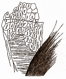
The position of this layer of cells is between the fenestrated membrane and the cortical scales; clear proof of this is obtained when the cortical scales happen to peel off from the shaft and adhere to the inner surface of the sheath. If the focus be adjusted to them, depressing it brings into view, 1st. the layer of nucleated cells; 2nd. the fenestrated membrane.
Subjoined is a drawing of the inner surface of a hair sheath, illustrating this.
Possibly it is the layer of cells here described which has been confounded by Kohlrausch and Krause with the fenestrated membrane, which has been described by Henle as consisting of anastomosing longitudinal fibres, and by Meyer (cited in Professor Henle's "Allgemeine Anatomie," p. 295), as a stage of development of the cortical scales.


|
THE
HUXLEY
FILE

|
| ||||||||||||||||||||||||||||||||||||||||||||||||||||||