
Contents
| Lesson I. | A General View of the Structure and Functions of the Human Body | 1 |
| Lesson II. | The Vascular System and the Circulation | 21 |
| Lesson III. | The Blood and the Lymph | 58 |
| Lesson IV. | Respiration | 75 |
| Lesson V. | The Sources of Loss and of Gain to the Blood | 101 |
| Lesson VI. | The Function of Alimentation | 133 |
| Lesson VII. | Motion and Locomotion | 156 |
| Lesson VIII. | Sensation and Sensory Organs | 187 |
| Lesson IX. | The Organ of Sight | 214 |
| Lesson X. | The Coalescence of Sensations with One Another and with Other States of Consciousness | 236 |
| Lesson XI. | The Nervous System and Innervation | 248 |
| Lesson XI. | Histology, or the Minute Structure of the Tissues | 272 |
| Appendix A. | Table of Anatomical and Physiological Constants | 297 |
| Appendix B. | Case of Mrs. A. | 302 |
[v]
A considerable number of illustrations have been added to this edition; and several of them have been taken, not from the Human subject, but from the Rabbit, the Sheep, the Dog, and the Frog, in order to aid those, who, in accordance with the recommendation contained in the Preface to the Second Edition, attempt to make their knowledge real, by acquiring some practical acquaintance with the facts of Anatomy and Physiology.
My thanks are again due to my friend Dr. Foster, F.R.S., for many valuable suggestions, and, more especially, for the trouble he has taken in superintending the execution of the new woodcuts.
London, September 1872.
[vii]
The present edition of the "Lessons in Elementary Physiology," has been very carefully revised. A few woodcuts have been added; others have been replaced by better ones, as in the case of the figures of the retina, which embody the results of Schultze's latest researches.
Some additions (but as few as possible, lest the book should insensibly lose its elementary character) have been made; among the most important I count the very useful "Table of Anatomical and Physiological Constants" drawn up for me by Dr. Michael Foster, for whose friendly aid I am again glad to express my thanks.
It will be well for those who attempt to study Elementary Physiology, to bear in mind the important truth that the knowledge of science which is attainable by mere reading, though infinitely better than ignorance is knowledge of a very different kind from that which arises from direct contact with fact; and that the worth of the pursuit of science as an intellectual discipline is almost lost by those who seek it only in books.
As the majority of the readers of these Lessons will assuredly have no opportunity of studying anatomy or physiology upon the human subject, these remarks may seem discouraging. But they are not so in reality. For the purpose of acquiring a practical, though elementary, acquaintance with physiological anatomy and histology, the organs and tissues of the commonest domestic animals afford ample materials. The principal points in the structure and mechanism of the heart, the lungs, the kidneys, or the eye, of man, may be perfectly illustrated by the corresponding parts of a sheep; while the phenomena of the circulation, and many of the most important properties of living tissues, are better shown by the common frog than by any of the higher animals.
Under these circumstances there really is no reason why the teaching of elementary physiology should not be made perfectly sound and thorough. But it should be remembered that, unless the learner has previously acquired a knowledge of the elements of Physics and of Chemistry, his path will be beset with difficulties and delays.
London, July 1868.
[ix]
The following "Lessons in Elementary Physiology" are primarily intended to serve the purpose of a textbook for teachers and learners in boys' and girls' schools.
My object in writing them has been to set down, in plain and concise language, that which any person who desires to become acquainted with the principles of Human Physiology may learn, with a fair prospect of having but little to unlearn as our knowledge widens.
It is only by inadvertence, or from an error in judgment, therefore, that the book contains any statement, or doctrine, which cannot be regarded as the common property of all physiologists. I have endeavoured simply to play the part of a sieve, and to separate the well-established and essential from the doubtful and the unimportant portions of the vast mass of knowledge and opinion we call Human Physiology.
The originals of the woodcuts are, for the most part, to be found in the works of Bourgery, Gray, Henle, and Kölliker. A few are new.
I am particularly indebted to my accomplished friend, Dr. Michael Foster, for the pains and trouble he has bestowed upon the Lessons in their passage through the press.
The Royal School of Mines, London,
October 1866.
Lesson III
[58]
1. In order to become properly acquainted with the characters of the blood it is necessary to examine it with a microscope magnifying at least three or four hundred diameters. Provided with this instrument, a hand lens, and some slips of thick and thin glass, the student will be enabled to follow the present Lesson.
The most convenient mode of obtaining small quantities of blood for examination is to twist a piece of string, pretty tightly, round the middle of the last joint of the middle, or ring finger, of the left hand. The end of the finger will immediately swell a little, and become darker coloured, in consequence of the obstruction to the return of the blood in the veins caused by the ligature. When in this condition, if it be slightly pricked with a sharp clean needle (an operation which causes hardly any pain), a good-sized drop of blood will at once exude. Let it be deposited on one of the slips of thick glass, so as to spread it out evenly into a thin layer. Let a second slide receive another drop, and, to keep it from drying, let it be put under an inverted watch-glass or wine-glass, with a bit of wet blotting-paper inside. Let a third drop be dealt with in the same way, a few granules of common salt being added to the drop.
2. To the naked eye the layer of blood upon the first slide will appear of a pale reddish colour, and quite clear and homogeneous. But on viewing it with even a pocket lens its apparent homogeneity will disappear, and it will [59] look like a mixture of excessively fine yellowish-red particles, like sand, or dust, with a watery, almost colourless, fluid. Immediately after the blood is drawn, the particles will appear to be scattered very evenly through the fluid, but by degrees they aggregate into minute patches, and the layer of blood becomes more or less spotty.
The "particles" are what are termed the corpuscles of the blood; the nearly colourless fluid in which they are suspended is the plasma.
The second slide may now be examined. The drop of blood will be unaltered in form, and may perhaps seem to have undergone no change. But if the slide be inclined, it will be found that the drop no longer flows; and, indeed, the slide may be inverted without the disturbance of the drop, which has become solidified, and may be removed, with the point of a penknife, as a gelatinous mass. The mass is quite soft and moist, so that this setting, or coagulation, of a drop of blood is something very different from its drying.
On the third slide, this process of coagulation will be found not to have taken place, the blood remaining as fluid as it was when it left the body. The salt, therefore, has prevented the coagulation of the blood. Thus this very simple investigation teaches that blood is composed of a nearly colourless plasma, in which many coloured corpuscles are suspended; that it has a remarkable power of coagulating; and that this coagulation may be prevented by artificial means, such as the addition of salt.
3. If, instead of using the hand lens, the drop of blood on the first slide be placed under the microscope, the particles, or corpuscles, of the blood will be found to be bodies with very definite characters, and of two kinds, called respectively the red corpuscles and the colourless corpuscles. The former are much more numerous than the latter, and have a yellowish-red tinge; while the latter, somewhat larger than the red corpuscles, are, as their name implies, pale and devoid of coloration.
4. The corpuscles differ also in other and more important respects. The red corpuscles (Fig. 17) are flattened circular disks, on an average 1/3200 of an inch in diameter, and having about one-fourth of that thickness. It follows that rather more than l0,000,000 of them will lie on a space [60] one inch square, and that the volume of each corpuscle does not exceed 1/120000000000 of a cubic inch.
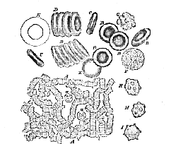
A. Moderately magnified. The red corpuscles are seen lying in rouleaux; at a and a are seen two white corpuscles.
B. Red corpuscles much more highly magnified, seen in face;
C. ditto, seen in profile;
D. ditto, in rouleaux rather more highly magnified;
E. a red corpuscle swollen into a sphere by inhibition of water.
F. A white corpuscle magnified same as B;
G. ditto, throwing out some blunt processes;
K. ditto, treated with ascetic acid, and showing nucleus magnified same as D.
H. Red corpuscles puckered or crenate all over.
I. Ditto, at the edge only.
The broad faces of the disks are not flat, but somewhat concave, as if they were pushed in towards one another. Hence the corpuscle is thinner in the middle than at the edges, and when viewed under the microscope, by transmitted light, looks clear in the middle and darker at the edges, or dark in the middle and clear at the [61] edges, according to circumstances. When, on the other hand, the disks roll over and present their edges to the eye, they look like rods. All these varieties of appearance may be made intelligible by turning a round biscuit or muffin, bodies similar in shape to the red corpuscles, in various ways before the eye.
The red corpuscles are very soft, flexible, and elastic bodies, so that they readily squeeze through apertures and passages narrower than their own diameters, and immediately resume their proper shapes (Fig. 16, G. H.). The exterior of each corpuscle is denser than its interior, which contains a semi-fluid, or quite fluid matter, of a red colour, called hæmoglobin. By proper processes this may be resolved into an albuminous substance sometimes called globulin, and a peculiar colouring matter, which is called hæmatin. The interior substance presents no distinct structure.
From the density of the outer as compared with the inner substance of each corpuscle, they are, practically, small flattened bags, or sacs, the form of which may be changed by altering the density of the plasma. Thus, if it be made denser by dissolving saline substances, or sugar, in it, water is drawn from the contents of the corpuscle to the dense plasma, and the corpuscle becomes still more flattened and very often much wrinkled. On the other hand, if the plasma be diluted with water, the latter forces itself into and dilutes the contents of the corpuscle, causing the latter to swell out, and even become spherical; and, by adding dense and weak solutions alternately, the corpuscles may be made to become successively spheroidal and discoidal. Exposure to carbonic acid gas seems to cause the corpuscles to swell out; oxygen gas, on the contrary, appears to flatten them.
5. The colourless corpuscles (Fig. 17, a a, F. G. K.) are larger than the red corpuscles, their average diameter being 1/2500 of an inch. They are further seen, at a glance, to differ from the red corpuscles by the extreme irregularity of their form, and by their tendency to attach themselves to the glass slide, while the red corpuscles float about and tumble freely over one another.
A still more remarkable feature of the colourless [62] corpuscles than the irregularity of their form is the unceasing variation of shape which they exhibit. The form of a red corpuscle is changed only by influences from without, such as pressure, or the like; that of the colourless corpuscle is undergoing constant alteration, as the result of changes taking place in its own substance. To see these changes well, a microscope with a magnifying power of five or six hundred diameters is requisite; and, even then, they are so gradual that the best way to ascertain their existence is to make a drawing of a given colourless corpuscle at intervals of a minute or two. This is what has been done with the corpuscle represented in Fig. 18, in which a represents the form of the corpuscle when first observed; b, its form a minute afterwards; c, that at the end of the second; d, that at the end of the third; and e, that at the end of the fifth minute.

(Magnified about 600 diameters.) The interval between the forms a , b, c, d, was a minute; between d and e two minutes; so that the whole series of changes from a to e took five minutes.
Careful watching of a colourless corpuscle, in fact, shows that every part of its surface is constantly changing–undergoing active contraction, or being passively dilated by the contraction of other parts. It exhibits contractility in its lowest and most primitive form.
6. While they are thus living and active, no correct notion can be formed of the structure of the colourless corpuscles. By diluting the blood with water, or, still better, with water acidulated with acetic acid, the corpuscles are killed, and become distended, so that their real nature is shown. They are then seen to be spheroidal bags, or sacs, with very thin walls; and to contain in their interior a fluid which is either clear or granular together with a spheroidal vesicular body, which is called [63] the nucleus (Fig. 17, K). It sometimes, though very rarely, happens that the nucleus has a red tint.
The sac-like colourless corpuscle, with its nucleus, is what is called a nucleated cell. It will be observed that it lives in a free state in the plasma of the blood, and that it exhibits an independent contractility. In fact, except that it is dependent for the conditions of its existence upon the plasma, it might be compared to one of those simple organisms which are met with in stagnant water, and are called Amœbæ.
7. That the red corpuscles are in some way or other derived from the colourless corpuscles may be regarded as certain. but the steps of the process have not been made out with perfect certainty. There is very great reason, however, for believing that the red corpuscle is simply the nucleus of the colourless corpuscle somewhat enlarged; flattened from side to side; changed, by development within its interior of a red colouring matter; and set free by the bursting of the sac or wall of the colourless corpuscle. Its other words, the red corpuscle is a free nucleus.
The origin of the colourless corpuscles themselves is not certainly determined; but it is highly probable that they are constituent cells of particular parts of the solid substance of the body which have been detached and carried into the blood, and that this process is chiefly effected in what are called the ductless glands (Lesson V. § 27), from whence the detached cells pass, as lymph-corpuscles, directly or indirectly, into the blood.
The following facts are of importance in their bearing on the relation between the different kinds of corpuscles:–
(a) The invertebrate animals1 which have true blood-corpuscles, possess only such as resemble the colourless corpuscles of man.
(b) The lowest vertebrate animal, the Lancelet (Amphioxus), possesses only colourless corpuscles; and the very young embryos2 of all vertebrate animals have only colourless and nucleated corpuscles.
[64] (c) All the vertebrated animals, the young of which are born from eggs,3 have two kinds of corpuscles–colourless corpuscles, like those of man, and large red-coloured corpuscles, which are generally oval, and further differ from those of man in presenting a nucleus. In fact, they are simply the colourless corpuscles enlarged and coloured.
[64] (d ) All animals which suckle their young (or what are called mammals) have, like man, two kinds of corpuscles; colourless ones, and small coloured corpuscles–the latter being always flattened, and devoid of any nucleus. They are usually circular, but in the camel tribe they are elliptical. And it is worthy of remark that, in these animals, the nuclei of the colourless corpuscles become elliptical.
(e) The colourless corpuscles differ much less from one another in size and form, in the vertebrate series, than the coloured. The latter are smallest in the little Musk Deer, in which animal they are about a quarter as large as those of a man. On the other hand, the red corpuscles are largest in the Amphibia (or Frogs and Salamanders), in some of which animals they are ten times as long as in man.
8. As the blood dies, its several constituents, which have now been described, undergo marked changes.
The colourless corpuscles lose their contractility, but otherwise undergo little alteration. They tend to cohere neither with one another, nor with the red corpuscles, but adhere to the glass plate on which they are placed.
It is quite otherwise with the red corpuscles which at first, as has been said, float about and roll, or slide, over each other quite freely. After a short time (the length of which varies in different persons, but usually amounts to two or three minutes), they seem, as it were, to become sticky, and tend to cohere; and this tendency increases until, at length, the great majority of them become applied face to face, so as to form long series, like rolls of coin. The end of one roll cohering with the sides of another, a network of various degrees of closeness is produced (Fig. 17, A.).
The corpuscles remain thus coherent for a certain length of time, but eventually separate and float freely [65] again. The addition of a little water, or dilute acids or saline solutions, will at once cause the rolls to break up.
It is from this running of the corpuscles together into patches of network that the change noted above in the appearances of the layer of blood, viewed with a lens, arises. So long as the corpuscles are separate, the sandy appearance lasts; but when they run together, the layer appears patchy or spotted.
The red corpuscles rarely, if ever, all run together into rolls, some always remaining free in the meshes of the net. In contact with air, or if subjected to pressure, many of the red corpuscles become covered with little knobs, so as to look like minute mulberries–an appearance which has been mistaken for a breaking up, or spontaneous division, of the corpuscles (Fig. 17, H. H.).
9. There is a still more remarkable change which the red blood-corpuscles occasionally undergo. Under certain circumstances, the peculiar red substance which forms the chief mass of their contents, and which has been called hæmoglobin (from its readily breaking up into globulin and hæmatin, § 6), separates in a crystalline form. In man, these crystals have the shape of prisms; in other animals they take other forms. Human blood crystallizes with difficulty, but that of the guinea-pig, rat, or dog much more easily. The best way to see these blood-crystals is to take a little rat's blood, from which the fibrin has been removed, shake it up with a little ether, and let it stand in the cold for some hours. A sediment will form at the bottom, which, when examined with the microscope, will be found to consist of long narrow crystals. Crystallization is much assisted by adding after the ether a small quantity of alcohol.
10. When the layer of blood has been drawn ten or fifteen minutes, the plasma will be seen to be no longer clear. It then exhibits multitudes of extremely delicate filaments of a substance called Fibrin, which have been deposited from it, and which traverse it in all directions, uniting with one another and with the corpuscles, and binding the whole into a semi-solid mass.
It is this deposition of fibrin which is the cause of the apparent solidification, or coagulation, of the drop upon the second slide; but the phenomena of coagula[66]tion, which are of very great importance, cannot be properly understood until the behaviour of the blood, when drawn in larger quantity than a drop, has been studied.
11. When, by the ordinary process of opening a vein with a lancet, a quantity of blood is collected into a basin, it is at first perfectly fluid: but in a very few minutes it becomes, through coagulation, a jelly-like mass, so solid that the basin may be turned upside down without any of the blood being spilt. At first the clot is a uniform red jelly, but very soon drops of a clear yellowish watery-looking fluid make their appearance on the surface of the clot, and on the sides of the basin. These drops increase in number, and run together, and after a while it has become apparent that the originally uniform jelly has separated into two very different constituents–the one a clear, yellowish liquid; the other a red, semi-solid mass, which lies in the liquid, and at the surface is paler in colour and firmer than in its deeper part.
The liquid is called the serum; the semi-solid mass the clot, or crassamentum. Now the clot obviously contains the corpuscles of the blood, bound together by some other substance; and this last, if a small part of the clot be examined microscopically, will be found to be that fibrous-looking matter, fibrin, which has been seen forming in the thin layer of blood. Thus the clot is equivalent to the corpuscles plus the fibrin of the plasma, while the serum is the plasma minus the fibrinous elements which it contained.
12. The corpuscles of the blood are slightly heavier than the plasma, and therefore, when the blood is drawn they sink very slowly towards the bottom. Hence the upper part of the clot contains fewer corpuscles, and is lighter in colour, than the lower part–there being fewer corpuscles left in the upper layer of plasma for the fibrin to catch when it sets. And there are some conditions of the blood in which the corpuscles run together much more rapidly and in denser masses than usual. Hence they more readily overcome the resistance of the plasma to their falling, just as feathers stuck together in masses fall much more rapidly through the air than the same feathers when loose. When this is the case, the [67] upper stratum of plasma is quite free from red corpuscles before the fibrin forms in it; and, consequently, the uppermost layer of the clot is nearly white: it receives the name of the buffy coat.
After the clot is formed, the fibrin shrinks and squeezes out much of the serum contained within its meshes; and, other things being equal, it contracts the more the fewer corpuscles there are in the way of its shrinking. Hence, when the buffy coat is formed, it usually contracts so much as to give the clot a cup-like upper surface.
Thus the buffy coat is fibrin naturally separated from the red corpuscles; the same separation may be effected, artificially, by whipping the blood with twigs as soon as it is drawn, until its coagulation is complete. Under these circumstances the fibrin will collect upon the twigs, and a red fluid will be left behind, consisting of the serum plus the red corpuscles, and many of the colourless ones.
13. The coagulation of the blood is hastened, retarded, or temporarily prevented by many circumstances.
(a) Temperature.–A high temperature accelerates the coagulation of the blood; a low one retards it very greatly; and some experimenters have stated that, when kept at a sufficiently low temperature, it does not coagulate at all.
(b) The addition of soluble matter to the blood.–Many saline substances, and more especially sulphate of soda and common salt, dissolved in the blood in sufficient quantity, prevent its coagulation; but coagulation sets in when water is added, so as to dilute the saline solution.
(c) Contact with living or not living matter.–Contact with not living matter promotes the coagulation of the blood. Thus, blood drawn into a basin begins to coagulate first where it is in contact with the sides of the basin; and a wire introduced into a living vein will become coated with fibrin, although perfectly fluid blood surrounds it.
On the other hand, direct contact with living matter retards, or altogether prevents, the coagulation of the blood, Thus blood remains fluid for a very long time in a portion of a vein which is tied at each end.
The heart of a turtle remains alive for a lengthened period (many hours or even days) after it is extracted from [68] the body; and, so long as it remains alive, the blood contained in it will not coagulate, though, if a portion of the same blood be removed from the heart, it will coagulate in a few minutes.
Blood taken from the body of the turtle, and kept from coagulating by cold for some time, may be poured into the separated, but still living, heart and then will not coagulate.
Freshly deposited fibrin acts somewhat like living matter, coagulable blood remaining fluid for a long time in tubes coated with such fibrin.
14. The coagulation of the blood is an altogether physico-chemical process, dependent upon the properties of certain of the constituents of the plasma, apart from the vitality of that fluid. This is proved by the fact that if blood-plasma be prevented from coagulating by cold, and greatly diluted, a current of carbonic acid passed through it will throw down a white powdery substance. If this white substance be dissolved in a weak solution of common salt, or in an extremely weak solution of potash or soda, it, after a while, coagulates, and yields a clot of true pure fibrin. It would be absurd to suppose that a substance which has been precipitated from its solution and redissolved, still remains alive.
There are reasons for believing that this white substance consists of two constituents of very similar composition, which exist separately in living blood, and the union of which is the cause of the act of coagulation. These reasons may be briefly stated thus:–The pericardium and other serous cavities in the body contain a clear fluid, which has exuded from the blood-vessels, and contains the elements of the blood without the blood-corpuscles. This fluid sometimes coagulates spontaneously, as the blood plasma would do, but very often shows no disposition to spontaneous coagulation. When this is the case, it may nevertheless be made to coagulate, and yield a true fibrinous clot, by adding to it a little serum of blood.
Now, if serum of blood be largely diluted with water and a current of carbonic be gas passed through it, a white powdery substance will be thrown down; this, redissolved in a dilute saline, or extremely dilute alkaline, [69] solution will, when added to the pericardial fluid, produce even as good a clot as that obtained with the original serum.
This white substance has been called globulin. It exists not only in serum, but also, though in smaller quantities, in connective tissue, in the cornea, in the humours of the eye, and in some other fluids of the body.
It possesses the same general chemical properties as the albuminous substance which enters so largely into the composition of the red corpuscles (§ 4), and hence, at present, bears the same name. But when treated with chemical reagents, even with such as do not produce any appreciable effect on its chemical composition, it very speedily loses its peculiar power of causing serous fluids to coagulate. For instance, this power is destroyed by an excess of alkali, or by the presence of acids.
Hence, though there is great reason to believe that the fibrino-plastic globulin (as it has been called) which exists in serum does really come from the red corpuscles, the globulin which is obtained in large quantities from these bodies, by the use of powerful reagents, has no coagulating effect at all on pericardial or other serous fluids.
Though globulin is so susceptible of change when in solution, it may be dried at a low temperature and kept in the form of powder for many months, without losing its coagulating power.
Thus globulin, added, under proper conditions, to serous effusion, is a coagulator of that effusion, giving rise to the development of fibrin in it.
It does so by its interaction with a substance contained in the serous effusion, which can be extracted by itself, and then plays just the same part towards a solution of globulin, as globulin does towards its solution. This substance has been called fibrinogen. It is exceedingly like globulin, and may be thrown down from serous exudation by carbonic acid, just as globulin may be precipitated from the serum of the blood. When redissolved in an alkaline solution, and added to any fluid containing globulin, it acts as a coagulator of that fluid, and gives rise to the development of a clot of fibrin in it. In accordance with what has just been stated, serum of blood which has completely coagulated may be kept in one [70] vessel, and pericardial fluid in another, for an indefinite period, if spontaneous decomposition be prevented, without the coagulation of either. But let them be mixed, and coagulation sets in.
Thus it seems to be clear, that the coagulation of the blood, and the formation of fibrin, are caused primarily by the interaction of two substances (or two modifications of the same substance), globulin or fibrinoplastin and fibrinogen, the former of which may be obtained from the serum of the blood, and from some tissues of the body; while the latter is known, at present, only in the plasma of the blood, of the lymph, and of the chyle, and in fluids derived from them.
15. The proverb that "blood is thicker than water" is literally true, as the blood is not only "thickened" by the corpuscles, of which it has been calculated that no fewer than 70,000,000,000 (eighty times the number of the human population of the globe) are contained in a cubic inch, but is rendered slightly viscid by the solid matters dissolved in the plasma. The blood is thus rendered heavier than water, its specific gravity being about 1055. In other words, twenty cubic inches of blood have about the same weight as twenty-one cubic inches of water.
The corpuscles are heavier than the plasma, and their volume is usually somewhat less than that of the plasma. Of colourless corpuscles there are usually not more than three or four for every thousand of red corpuscles; but the number varies very much, increasing shortly after food is taken, and diminishing in the intervals between meals.
The blood is hot, being about 100° Fahrenheit.
16. Considered chemically, the blood is an alkaline fluid, consisting of water, of solid and of gaseous matters.
The proportion of these several constituents vary according to age, sex, and condition, but the following statement holds good on the average:–
In every 100 pairs of the blood there are 79 parts of water and 21 parts of dry solids; in other words, the water and the solids of the blood stand to one another in about the same proportion as the nitrogen and the oxygen of the air. Roughly speaking, one quarter of the blood [71] is dry, solid matter; three quarters water. Of the 21 parts of dry solids, 12 (= 4/7ths) belong to the corpuscles. The remaining 9 are about two-thirds (6.7 parts = 2/7ths) albumin (a substance like white of egg, coagulating by heat), and one-third (= 1/7th of the whole solid matter) a mixture of saline, fatty, and saccharine matters, sundry products of the waste of the body, and fibrin. The quantity of the latter constituent is remarkably small in relation to the conspicuous part it plays in the act of coagulation. Healthy blood, in fact, yields in coagulating not more than from two to four parts in a thousand of its weight of fibrin.
The total quantity of gaseous matter contained in the blood is equal to rather less than half the volume of the blood; that is to say, 100 cubic inches of blood will contain rather less than 50 cubic inches of gases. These gaseous matters are carbonic acid, oxygen, and nitrogen; or, in other words, the same gases as those which exist in the atmosphere, but in totally different proportions; for whereas air contains nearly three-fourths nitrogen, one-fourth oxygen, and a mere trace of carbonic acid, the average composition of the blood gases is nearly two-thirds carbonic acid, rather less than one-third oxygen, and not one-tenth nitrogen.
It is important to observe that blood contains much more oxygen gas than could be held in solution by pure water at the same temperature and pressure. This power of holding oxygen appears in some way to depend upon the corpuscles, firstly, because mere serum has no greater power of absorbing oxygen than pure water has; and secondly, because red corpuscles suspended in water instead of serum absorb oxygen very readily. The oxygen thus held by the red corpuscles is readily given up by them for purposes of oxidation, and indeed can be removed from them by means of a mercurial gas pump. It would appear that the connection between the oxygen and the red corpuscles is of a peculiar nature, being a sort of loose chemical combination with one of their constituents, that constituent being the hæmoglobin; for solutions of hæmoglobin behave towards oxygen exactly as blood does.
The corpuscles differ chemically from the plasma, in [72] containing a large proportion of the fats and phosphates, all the iron, and almost all the potash, of the blood; while the plasma, on the other hand, contains by far the greater part of the chlorine and the soda.
17. The blood of adults contains a larger proportion of solid constituents than that of children, and that of men more than that of women; but the difference of sex is hardly at all exhibited by persons of flabby, or what is called lymphatic, constitution.
Animal diet tends to increase the quantity of the red corpuscles; a vegetable diet and abstinence to diminish them. Bleeding exercises the same influence in a still more marked degree, the quantity of red corpuscles being diminished thereby in a much greater proportion than that of the other solid constituents of the blood
18. The total quantity of blood contained in the body varies at different times, and the precise ascertainment of its amount is very difficult. It may probably be estimated, on the average, at not less than one-thirteenth of the weight of the body.
19. The function of the blood is to supply nourishment to, and take away waste matters from, all parts of the body. It is absolutely essential to the life of every part of the body that it should be in such relation with a current of blood, that matters can pass freely from the blood to it, and from it to the blood, by transudation through the walls of the vessels in which the blood is contained. And this vivifying influence depends upon the corpuscles of the blood. The proof of these statements lies in the following experiments: If the vessels of a limb of a living animal be tied in such a manner as to cut off the supply of blood from the limb, without affecting it in any other way, all the symptoms of death will set in. The limb will grow pale and cold, it will lose its sensibility, and volition will no longer have power over it; it will stiffen, and eventually mortify and decompose.
But, even when the death stiffening has begun to set in, if the ligatures be removed, and the blood be allowed to flow into the limb, the stiffening speedily ceases, the temperature of the part rises, the sensibility of the skin returns, the will regains power over the muscles, and, in short, the part returns to its normal condition.
[73] If, instead of simply allowing the blood of the animal operated upon to flow again, such blood, deprived of its fibrin by whipping, but containing its corpuscles, be artificially passed through the vessels, it will be found as effectual a restorative as entire blood; while, on the other hand, the serum (which is equivalent to whipped blood without its corpuscles) has no such effect
It is not necessary that the blood thus artificially injected should be that of the subject of the experiment. Men, or dogs, bled to apparent death, may be at once and effectually revived by filling their veins with blood taken from another man, or dog; an operation which is known by the name of transfusion.
Nor is it absolutely necessary for the success of this operation that the blood used in transfusion should belong to an animal of the same species. The blood of a horse will permanently revive an ass, and, speaking generally, the blood of one animal may be replaced without injurious effects by that of another closely-allied species; while that of a very different animal will be more or less injurious, and may even cause immediate death.
20. The Lymph, which fills the lymphatic vessels, is, like the blood, an alkaline fluid, consisting of a plasma and corpuscles, and coagulates by the separation of fibrin from the plasma. The lymph differs from the blood in its corpuscles being all of the colourless kind, and in the very small proportion of its solid constituents, which amount to only about 5 per cent. of its weight. Lymph may, in fact, be regarded as blood minis its red corpuscles, and diluted with water, so as to be somewhat less dense than the serum of blood, which contains about 8 per cent. of solid matters.
A quantity of fluid equal to that of the blood is probably poured into the blood, daily, from the lymphatic system. This fluid is in great measure the mere overflow of the blood itself–plasma which has exuded from the capillaries into the tissues, and which has not been taken up again into the venous current; the rest is due to the absorption of chyle from the alimentary canal.
1 Invertebrate animals are animals devoid of backbones, such as insects, snails, sea-anemones, &c. Vertebrate animals are fishes, amphibia, reptiles, birds, and mammals. 2 An embryo is the rudimentary unborn young of any creature. 3 These are fishes, amphibia, reptiles, and birds.
[187]
SENSATIONS AND SENSORY ORGANS.
1. The agent by which all the motor organs (except the cilia) described in the preceding Lesson are set at work, is muscular fibre. But, in the living body, muscular fibre is made to contract only by a change which takes place in the motor or efferent nerve, which is distributed to it. This change again is effected only by the activity of the central nervous organ, with which the motor nerve is connected. The central organ is thrown into activity immediately, or ultimately, only by the influence of changes which take place in the molecular condition of nerves, called sensory or afferent, which are connected, on the one hand, with the central organ, and, on the other hand, with some other part of the body. Finally, the alteration of the afferent nerve is itself produced only by changes in the condition of the part of the body with which it is connected; which changes usually result from external impressions.
2. Thus the great majority (if not the whole) of the movements of the body and of its parts, are the effect of an influence (technically termed a stimulus or irritation) applied directly, or indirectly, to the ends of afferent nerves, and giving rise to a molecular change, which is propagated along their substance to the central nervous organ with which they are connected. The molecular activity of the afferent nerve communicates itself to the central organ, and is then transmitted along the motor nerves, which pass from the central organ to the muscles affected. And, when the disturbance in the molecular [188] condition of the efferent nerves reaches their extremities, it is communicated to the muscular fibres, and causes their particles to take up a new position, so that each fibre shortens and becomes thicker.
3. Such a series of molecular changes as that just described is called a reflex action–the disturbance caused by the irritation being as it were reflected back, along the motor nerves, to the muscles.
A reflex action, strictly so called, takes place without our knowing anything about it, and hundreds of such actions are going on continually in our bodies without our being aware of them. But it very frequently happens that we learn that something is going on, when a stimulus affects our afferent nerves, by having what we call a feeling or sensation. We class sensations along with emotions, and volitions, and thoughts, under the common head of states of consciousness. But what consciousness is, we know not; and how it is that anything so remarkable as a state of consciousness comes about as the result of irritating nervous tissue, is just as unaccountable as any other ultimate fact of nature.
4. Sensations are of very various degrees of definiteness. Some arise within ourselves, we know not how or where, and remain vague and undefinable. Such are the sensations of uncomfortableness, or faintness, of fatigue, or of restlessness. We cannot assign any particular place to these sensations, which are very probably the result of affections of the afferent nerves in general brought about by the state of the blood, or that of the tissues in which they are distributed. And however real these sensations may be, and however largely they enter into the sum of our pleasures and pains, they tell us absolutely nothing of the external world. They are not only diffuse, but they are also subjective sensations.
5. What is termed the muscular sense is less vaguely localized than the preceding, though its place is still incapable of being very accurately defined. This muscular sensation is the feeling of resistance which arises when any kind of obstacle is opposed to the movement of the body, or of any part of it; and it is something quite different from the feeling of contact or even of pressure.
[189] Lay one hand flat on its back upon a table, and rest a disc of cardboard a couple of inches in diameter upon the ends of the outstretched fingers; the only result will be a sensation of contact–the pressure of so light a body being inappreciable. But put a two-pound weight upon the cardboard, and the sensation of contact will be accompanied, or even obscured, by the very different feeling of pressure. Up to this moment the fingers and arm have rested upon the table; but now let the hand be raised from the table, and another new feeling will make its appearance–that of resistance to effort. This feeling comes into existence with the exertion of the muscles which raise the arm, and is the consciousness of that exertion given to us by the muscular sense.
Anyone who raises or carries a weight, knows well enough that he has this sensation; but he may be greatly puzzled to say where he has it. Nevertheless, the sense itself is very delicate, and enables us to form tolerably accurate judgments of the relative intensity of resistances. Persons who deal in articles sold by weight, are constantly enabled to form very precise estimates of the weight of such articles by balancing them in their hands; and in this case, they depend in a great measure upon the muscular sense.
6. In a third group of sensations, each feeling, as it arises, is assigned to a definite part of the body, and is produced by a stimulus applied to that part of the body; but the bodies, or forces, which are competent to act as stimuli, are very various in character. Such are the sensations of touch, which is restricted to the integument covering the surface, and to some portions of the membranes lining the internal cavities of the body; and of taste and smell, which are similarly confined to certain regions of the mucous membrane of the mouth and nasal cavities.
Any portion of the body to which a sensation is thus restricted is called a sensory organ.
And lastly, in a fourth group of sensations, each feeling requires for its production the application of a single kind of stimulus to a very specially modified part of the integument. The latter serves as an intermediator between the physical agent of the sensation and the sensory nerve, [190] which is to convey to the brain the impulse necessary to awake in it that state of consciousness which we call the sensation. Such are the sensations of sight and hearing. The physical agents which can alone awaken these sensations (under natural circumstances) are light and sound. The modified parts of the integument, which alone are competent to intermediate between these agents and the nerves of sight and hearing, are the eye and the ear.
7. In every sensory organ it is necessary to distinguish the terminal expansion of the afferent or sensory nerve, and the structures which intermediate between this expansion and the physical agent which gives rise to the sensation.
And in each group of special sensations there are certain phenomena which arise out of the structure of the organ, and others which result from the operation of the central apparatus of the nervous system upon the materials supplied to it by the sensory organ.
8. The sense of Touch (including that of heat and cold) is possessed, more or less acutely, by all parts of the free surface of the body, and by the walls of the mouth and nasal passages.
Whatever part possesses this sense consists of a membrane (integumentary or mucous) composed of a deep layer made up of fibrous tissue, containing a capillary network and the ultimate terminations of the sensory nerves; and of a superficial layer consisting of epithelial or epidermic cells, among which are no vessels.
Wherever the sense of touch is delicate, the deep layer is not a mere flat expansion, but is raised up into multitudes of small, close-set, conical elevations (see Fig. 32), which are called papillæ. In the skin, the coat of epithelial or epidermic cells does not follow the contour of these papillæ, but dips down between them and forms a tolerably even coat over them. Thus, the points of the papillæ are much nearer the surface than the general plane of the deep layer whence these papillæ proceed.
Loops of vessels enter the papillæ, and the fine ultimate terminations of the sensory nerve-fibres distributed to the skin terminate in them, but in what way has not been thoroughly made out.
[191] In certain cases, the delicate fibrous sheath, or neurilemma, of the nerve which enters the papilla, enlarges in the papilla into an oval swelling, which is called a tactile corpuscle (see Lesson XII.). These corpuscles are found in the papillæ of those localities which are endowed with a very delicate sense of touch, as in the tips of the fingers, the point of the tongue, &c.
9. It is obvious, from what has been said, that no direct contact takes place between a body which is touched and the sensory nerve,–a thicker or thinner layer of epithelium, or epidermis, being situated between the two. In fact, if this layer is removed, as when a surface of the skin has been blistered, contact with the raw surface gives rise to a sense of pain, not to one of touch properly so called. Thus, in touch, it is the epidermis, or epithelium, which is the intermediator between the nerve and the physical agent, the external pressure being transmitted through the horny cells to the subjacent ends of the nerves, and the kind of impulse thus transmitted must be modified by the thickness and character of the cellular layer, no less than by the forms and number of the papillae.
10. Certain very curious phenomena appertaining to the sense of touch, are probably due to these varying anatomical arrangements. Not only is tactile sensibility to a single impression much duller in some parts than in others–a circumstance which might be readily accounted for by the different thickness of the epidermic layer–but the power of distinguishing double simultaneous impressions is very different. Thus, if the ends of a pair of compasses (which should be blunted with pointed pieces of cork) are separated by only one-tenth or one-twelfth of an inch, they will be distinctly felt as two, if applied to the tips of the fingers; whereas, if applied to the back of the hand in the same way, only one impression will be felt; and, on the arm, they may be separated for a quarter of an inch, and still only one impression will be perceived.
Accurate experiments have been made in different parts of the body, and it has been found that two points can be distinguished by the tongue, if only one-twenty-fourth of an inch apart; by the tips of the fingers if [192] one-twelfth of an inch distant; while they may be one inch distant on the cheek, and even three inches on the back, and still give rise to only one sensation.
11. The feeling of warmth, or cold, is the result of an excitation of sensory nerves distributed to the skin which are probably distinct from those which give rise to the sense of touch. And it would appear that the heat must be transmitted through the epidermic or epithelial layer, to give rise to this sensation; for, just as touching a naked nerve, or the trunk of a nerve, gives rise only to pain, so heating or cooling an exposed nerve, or the trunk of a nerve, gives rise not to a sensation of heat or cold, but simply to pain.
Again, the sensation of heat, or cold, is relative rather than absolute. Suppose three basins be prepared, one filled with ice-cold water, one with water as hot as can be borne, and the third with a mixture of the two. If the hand be put into the hot-water basin, and then transferred to the mixture, the latter will feel cold; but if the hand be kept awhile in the ice-cold water, and then transferred to the very same mixture, it will feel warm.
Like the sense of touch, the sense of warmth varies in delicacy in different parts of the body.
The cheeks are very sensitive, more so than the lips; the palms of the hands are more sensitive to heat than their backs. Hence a washerwoman holds her flat-iron to her cheek to test the temperature, and one who is cold spreads the palms of his hands to the fire.
12. The organ of the sense of Taste is the mucous membrane which covers the tongue, especially its back part, and the hinder part of the palate. Like that of the skin, the deep, or vascular, layer of the mucous membrane of the tongue is raised up into papillæ, but there are large, separate, and have separate coats of epithelium. Towards the tip of the tongue they are for the most part elongated and pointed, and are called filiform ; over the rest of the surface of the tongue, these are mixed with other larger papillæ, with broad ends and narrow bases, called fungiform ; but towards its root here are a number of large papillæ, arranged in the figure of a V with its point backwards, each of which [193] is like a fungiform papilla surrounded by a wall. These are the circumvallate papillæ (Fig. 61, C.p.). The larger of these papilla have subordinate small ones upon their surfaces. They are very vascular, and they receive nervous filaments from two sources, the one the nerve called glossopharyngeal, the other the gustatory, which is a branch of the fifth nerve.
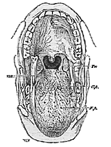
Uv. the uvula: Tn. the tonsil between the anterior and posterior pillars of the fauces; C. p. circumvallate papillæ, F.p. fungiform papillæ. The minute filiform papillæ cover the interspaces between these. On the right side the tongue is partially dissected to show the course of the filaments of the glossopharyngeal nerve, VIII.
The latter chiefly supplies the front of the tongue, the former its back and the adjacent part of the palate: and there [194] is reason to believe that it is the latter region which is more especially the seat of taste.
The great majority of the sensations we call taste, however, are in reality complex sensations, into which smell and even touch largely enter. When the sense of smell is interfered with, as when the nose is held tightly pinched, it is very difficult to distinguish the taste of various objects. An onion, for instance, the eyes being shut, may then easily be confounded with an apple.
13. The organ of the sense of Smell is the delicate mucous membrane which lines a part of the nasal cavities, and is distinguished from the rest of the mucous membrane of these cavities firstly, by possessing no cilia; secondly, by receiving its nervous supply from the olfactory, or first, pair of cerebral nerves, and not, like the rest of the mucous membrane, from the fifth pair.
Each nostril leads into a spacious nasal chamber, separated, in the middle line, from its fellow of the other side, by a partition, or septum, formed partly by cartilage and partly by bone, and continuous with that partition which separates the two nostrils one from the other. Below, each nasal chamber is separated from the cavity of the mouth by a floor, the bony palate (Figs. 62 and 63); and when this bony palate comes to an end, the partition is continued down to the root of the tongue by a fleshy curtain, the soft palate, which has been already described. The soft palate and the root of the tongue together, constitute, under ordinary circumstances, a moveable partition between the mouth and the pharynx, and it will be observed that the opening of the larynx, the glottis, lies behind the partition; so that when the root of the tongue is applied close to the soft palate no passage of air can take place between the mouth and the pharynx. But in the upper part of the pharynx above the partition are the two hinder openings of the nasal cavities (which are called the posterior nares) separated by the termination of the septum; and through these wide openings the air passes, with great readiness, from the nostrils along the lower part of each nasal chamber to the glottis, or in the opposite direction. It is by means of the passages thus freely open to the air that we breathe, as we ordinarily do, with the mouth shut.
Each nasal chamber rises, as a high vault, far above the
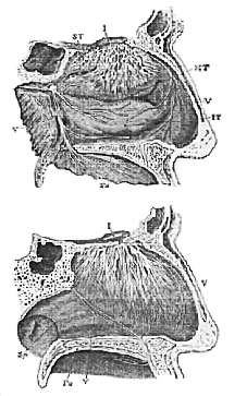
The upper figure represents the outer wall of the left nasal cavity; the lower figure the right side of the middle partition, or septum (Sp.), of the nose, which forms the inner wall of the right nasal cavity. I, the olfactory nerve and its branches; V, branches of the fifth nerve; Pa. the palate, which separates the nasal cavity from that of the mouth; S. T. the superior turbinal bone; M. T. the middle turbinal; I. T. the inferior turbinal. The letter I is placed in the cerebral cavity; and the partition on which the olfactory lobe rests, and through which the filaments of the olfactory nerves pass, is the cribriform plate.
[196] level of the arch of the posterior nares–in fact, about as high as the depression of the root of the nose. The uppermost and front part of its roof, between the eyes, is formed by a delicate horizontal plate of bone, perforated like a sieve by a great many small holes, and thence called the cribriform plate (Fig. 63, Cr.). It is this plate (with the membranous structures which line its two faces) alone which, in this region, separates the cavity of the nose from that which contains the brain. The olfactory lobes which are directly connected with, and form indeed a part of, the brain, enlarge at their ends, and their broad extremities rest upon the upper side of the cribriform plate; sending immense numbers of delicate filaments, the olfactory nerves, through it to the olfactory mucous membrane (Fig. 62).
On each wall of the septum this mucous membrane forms a flat expansion, but on the side walls of each nasal cavity it follows the elevations and depressions of the inner surfaces of what are called the upper and middle turbinal, or spongy bones. These bones are called spongy because the interior of each is occupied by air cavities separated from each other by very delicate partitions only and communicating with the nasal cavities. Hence the bones, though massive-looking, are really exceedingly light and delicate, and fully deserve the appellation of spongy (Fig. 63).
There is a third light scroll-like bone distinct from these two, and attached to the maxillary bone, which is called the inferior turbinal, as it lies lower than the other two, and imperfectly separates the air passages from the proper olfactory chamber (Fig. 62). It is covered by the ordinary ciliated mucous membrane of the nasal passage, and receives no filaments from the olfactory nerve (Fig. 62).
14. From the arrangements which have been described, it is clear that, under ordinary circumstances, the gentle inspiratory and expiratory currents will flow along the comparatively wide, direct passages afforded by so much of the nasal chamber as lies below the middle turbinal; and that they will hardly move the air enclosed in the narrow interspace between the septum and the upper and middle spongy bones, which is the proper olfactory chamber.
If the air currents are laden with particles of odorous [197] matter, they can only reach the olfactory membrane by diffusing themselves into this narrow interspace; and, if there be but few of these particles, they will run the risk of not reaching the olfactory mucous membrane at all, unless the air in contact with it be exchanged for some of the odoriferous air. Hence it is that, when we wish to perceive a faint odour more distinctly, we sniff, or snuff up the air. Each sniff is a sudden inspiration, the effect of which must reach the air in the olfactory chamber at the same time as, or even before, it affects that at the nostrils; and thus must tend to draw a little air out of that chamber from behind. At the same time, or immediately afterwards, the air sucked in at the nostrils entering with a sudden vertical rush, part of it must tend to flow directly into the olfactory chamber, and replace that thus drawn out.
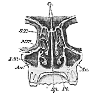
Fig. 63.–A transverse and Vertical Section of the Osseous Walls of the Nasal Cavity taken nearly through the Letter I in the foregoing Figure.
Cr. the cribriform Plate; S. T., M. T. the chambered superior and middle turbinal bones on which and on the septum (Sp.) the filaments of the olfactory nerve are distributed; I. T. the inferior turbinal bone; Pl.. the palate; An. the Anthrum or chamber which occupies the greater part of the maxillary bone and opens into the nasal cavity.
The loss of smell which takes place in the course of a severe cold may, in part, be due to the swollen state of [198] the mucous membrane which covers the inferior turbinal bones, which thus impedes the passage of odoriferous air to the olfactory chamber.
15. The Ear, or organ of the sense of Hearing, is very much more complex than either of the sensory organs yet described. It will be useful to distinguish the essential parts of this complicated apparatus from certain other parts, which, though of great assistance to the sense, are not absolutely necessary, and therefore may be called accessory.
The essential parts, on either side of the head, consist, substantially, of two peculiarly formed membranous bags, called, respectively, the membranous labyrinth and the scala media of the cochlea. Both these bags are lodged in cavities which they do not completely fill, situated in the midst of a dense and solid mass of bone (from its hardness called petrosal ), which forms a part of the temporal bone, and enters into the base of the skull.
Each bag is filled with a fluid, and is also supported in a fluid which fills the cavity in which it is lodged. In the interior of each bag, certain small, mobile, hard bodies are contained; and the ultimate filaments of the auditory nerves are so distributed upon the walls of the bags that their terminations must be knocked by the vibrations of these small hard bodies, should anything set them in motion. It is also quite possible that the vibrations of the fluid contents of the sacs may themselves suffice to affect the filaments of the auditory nerve; but, however this may be, any such effect must be greatly intensified by the cooperation of the solid particles.
In bathing in a tolerably smooth sea, on a rocky shore, the movement of the little waves as they run backwards and forwards is hardly felt by anyone lying down; but in bathing on a sandy and gravelly beach, the pelting of the showers of little stones and sand, which are raised and let fall by each wavelet, makes a very definite impression on the nerves of the skin.
Now, the membrane on which the ends of the auditory nerves are spread out is virtually a sensitive beach, and waves, which by themselves would not be felt, are readily perceived when they raise and let fall hard particles.
Both these membranous bags are lined by an epithelium.
[199] The auditory nerve after passing through the dense bone of the skull is distributed to certain regions of each bag, where its ultimate filaments come into peculiar connection with the epithelial lining. The epithelium itself too at these spots becomes specially modified. In certain parts of the membranous labyrinth, for instance, the epithelium connected with the terminations of the auditory nerve is produced into long, stiff, slender, hairlike processes (Fig. 64, d), which project into the fluid filling the bag, and which therefore are readily affected by any vibration of that fluid, and communicate the impulse to the ends of the nerves.
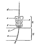
I. The epithelium of the ampulla. II. The membranous wall of the ampulla on which the epithelium rests. a, a filament of the auditory nerve running through the wall of the ampulla and breaking up into a fine network (b) in the epithelium; c, epithelium cell with long stiff hair-like filament, d (this cell is supposed by some to be directly continuous with the nerve network); e, cells, not bearing filaments, placed by the side of, and supporting the filament-bearing cells; f, a deeper layer of smaller cells.
In certain other parts of the same labyrinth these hairs are scanty or absent, but their place is supplied by minute angular particles of calcareous sand (called otoconia or otolithes), lying free in the fluid of the bag. [200] These, driven by the vibrations of that fluid, strike the epithelium and so affect the auditory nerve.
In the scala media of the cochlea, minute, rod-like bodies, called the fibres of Corti, and which are peculiarly modified cells of the epithelial lining of the scala, appear to serve the same object.
16. For simplicity's sake, the membranous labyrinth and the scala media have hitherto been spoken of as if they were simple bags; but this is not the case, each bag having a very curious and somewhat complicated form. (Figs. 65 and 66.)
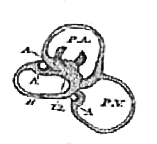
Ut. the Utriculus, or part of the vestibular sac, into which the semicircular canals open; A, A, A, the ampullæ; P.A. anterior vertical semicircular canal; P. V. posterior vertical semicircular canal; H. horizontal semicircular canal. The sacculus is not seen, as in the position in which the labyrinth Is drawn the sacculus lies behind the utriculus. The white circles on the ampullæ of the posterior vertical and horizontal canals indicate the cut ends of the branches of the auditory nerve ending in those ampullæ; the branches to the ampulla of the anterior vertical canal are seen in the space embraced by the canal, as is also the branch to the utriculus.
This form is also followed to a certain extent by the bony casing of the cavity in which each is lodged. Thus the membranous labyrinth is surrounded by a bony labyrinth, and the scala media is only a part of an intricate structure called the cochlea. The bony labyrinth and cochlea with all the parts inside each constitute together what is called the internal ear.
The membranous labyrinth (Fig. 65) has the figure of an oval vestibular sac, consisting of two parts, the one called utriculus, the other sacculus hemisphericus. The hoop-like semicircular canals open into the utriculus. They are three in number, and, two being vertical, are called the [201] anterior (P.A.) and posterior (P. V.) vertical semicircular canals; while the third, lying outside, and horizontally, is termed the external horizontal semicircular canal (H). One end of each of these canals is dilated into what is called an ampulla (A).
It is upon the walls of these ampullæ and those of the vestibular sac that the branches of the auditory nerve are distributed.
In each ampulla the nervous filaments may be traced to a transverse ridge caused by a thickening of the connective tissue which forms the walls of the canal (as well as of all other parts of the membranous labyrinth), and also by a thickening of the epithelium. Some of the epithelium cells are here prolonged into the fine hair-like processes described above. It is probable that these cells are specially connected with the terminations of the nerve filaments.
In the vestibule are similar but less marked ridges, or patches; here, however, the hair-like prolongations of the epithelium cells are absent or scanty, but, instead, otolithes are found in the fluid.
The fluid which fills the cavities of the semicircular canals and utriculus is termed endolymph. That which separates these delicate structures from the bony chambers in which they are contained is the perilymph. Each of these fluids is little more than water.
17. In the scala media1 the cochlea the primitive bag is drawn out into a long tube, which is coiled two and a half times on itself into a conical spiral, and lies in a much wider chamber of corresponding form, excavated in the petrous bone in such a way as to leave a central column of bony matter called the modiolus. The scala media has a triangular transverse section (Fig. 66), being bounded above and below by the membranous walls which converge internally and diverge externally. At their convergence, the walls are fastened to the edge of a thin plate of bone, the lamina spiralis (L.S. Fig. 66), which winds round the modiolus. At their divergence they are [202] fixed to the wall of the containing bony chamber, which thus becomes divided into two passages, communicating at the summit of the spire, but elsewhere separate. These two passages are called respectively the scala tympani and scala vestibuli, and are filled with perilymph.
The scala media, which thus lies between the other two scalæ, opens below, or at the broad end of the cochlea, by a narrow duct into the sacculus hemisphericus, but at its opposite end terminates blindly. (Fig. 70.)
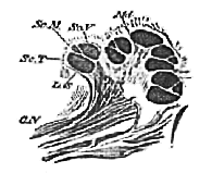
Sc.M. scala media; Sc. V. scala vestibuli; Sc. T. scala tympani; L. S. lamina spiralis; Md. bony axis, or modiolus, round which the scalæ are wound; C. N. cochlear nerve.
That branch of the auditory nerve which goes to supply the cochlea, enters the broad base of the central column or modiolus, and there divides into branches, which, spreading out in a spiral fashion in channels excavated in the bony tissue, are distributed to the lamina spiralis throughout its whole length. They do not end here, but in any section of the lamina spiralis (Fig. 66, L.S.) they may be found running outwards from the central column across the lamina towards the angle of the scala media, in which indeed they become finally lost.
The upper wall of the scala media, that which separates it from the scala vestibuli, is called the membrane of Reissner. The opposite or lower wall, which separates it from the scala tympani, is the basilar membrane. The latter is very elastic, and on it rest the fibres of Corti (C C, Fig. 67), each of which is composed of two filaments
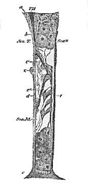
Fig. 67.–A Section through that Wall of the "Scala Media" of the Cochlea which lies next to the Scala Tympani.
a, That end of the lamina spiralis which passes into the inner wall, pillar, or mediolus of the bony cochlea; c, the outer wall of the bony cochlea; Sca. T the cavity of the scala tympani; Sca. M. the cavity of the scala media: d the elastic basilar membrane which separates the scala media from the scala tympani; V. a vessel which lies in this, cut through; c. the so-called mem[204]brane of Corti; C C', the fibres of Corti; VII. the filaments of the auditory nerve. It is doubtful whether the membrane of Corti really has the extent and connections given to it in this figure, which must not be taken for more than a general representation of the disposition of the parts. The membrane of Reissner, which separates the scala media from the scale vestibuli, is not represented.
[204] joined at an angle. An immense number of these filaments are set side by side, with great regularity, throughout the whole length of the scala media, so that this organ presents almost the appearance of a key-board, if viewed from either the scala vestibuli or the scala tympani. These fibres of Corti lie among a number of epithelium cells forming the lining of the scala media at this part, and those cells which are close to the fibres of Corti have a peculiarly modified form. The ends of the nerves have not yet been distinctly traced, but they probably come into close relation either with these fibres or with the modified epithelium cells lying close to them, which are capable of being agitated by the slightest impulse.
18. These essential parts of the organ of hearing are, we have seen, lodged in chambers of the petrous part of the temporal bone. Thus the membranous labyrinth is contained in a bony labyrinth of corresponding form, of which that part which lodges the sac is termed the vestibule, and those portions which contain the semicircular canals, the bony semicircular canals. And the scala media is contained in a spirally-coiled chamber, the cochlea, which it divides into two passages. Of these, one, the scala vestibuli, is so called because at the broad end or base of the cochlea it opens directly by a wide aperture into the vestibule; by this opening the perilymph which fills the vestibule and bony semicircular canals and surrounds the membranous labyrinth, is put in free communication with the perilymph which fills the scala vestibuli of the cochlea, and, by means of the communication which exists between the two scalæ at the summit of the spire, with that of the scala tympani also.
In the fresh state, this collection of chambers in the petrous bone is perfectly closed; but in the dry skull there are two wide openings, termed fenestræ, or windows, on its outer wall; i.e. on the side nearest the outside of the skull. Of these fenestræ, one, termed ovalis (the [205] oval window), is situated in the wall of the vestibular cavity, the other, rotunda (the round window), behind and below this, is the open end of the scala tympani at the base of the spire of the cochlea. In the fresh state, each of these windows or fenestræ is closed by a fibrous membrane, continuous with the periosteum of the bone.
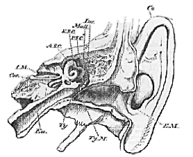
Fig. 68.–Transverse Section through the Side Walls of the Skull to show the Parts of Ear.
Co. Concha or external ear: E.M. external auditory meatus: Ty. M. tympanic membrane; Inc. Mall. incus and malleus; A.S.C. P.S.C. E.S.C. anterior, posterior and external semicircular canals; Coc. cochlea; Eu. Eustachian tube; I.M. internal auditory meatus, through which the auditory nerve passes to the organ of hearing.
The fenestra rotunda is closed only by membrane; but fastened to the centre of the membrane of the fenestra ovalis, so as to leave only a narrow margin, is an oval plate of bone, part of one of the little bones to be described shortly.
19. The outer wall of the internal ear is still far away from the exterior of the skull. Between it and the visible opening of the ear, in fact, are placed in a straight line, [206] first the drum of the ear, or tympanum ; secondly, the long external passage, or meatus (Fig. 68).
The drum of the ear and the external meatus, which together constitute the middle ear, would form one cavity, were it not that a delicate membrane, the tympanic membrane (Ty.M. Fig. 68), is tightly stretched in an oblique direction across the passage, so as to divide the comparatively small cavity of the drum from the meatus.
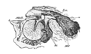
Fig. 69.–The Membrane of the drum of the Ear seen from the inner Side, with the small Bones of the Ear; and the Walls of the Tympanum, with the Air-cells in the Mastoid Part of the Temporal Bone.
M.C. mastoid cells; Mall. malleus; Inc. Incus; St. stapes; a b, lines drawn through the horizontal axis on which the malleus and the incus turn.
The membrane of the tympanum thus prevents any communication by means of the meatus, between the drum and the external air, but such a communication is provided, though in a roundabout way, by the Eustachian tube (Eu. Fig. 68), which leads directly from the fore part of the drum inwards to the roof of the pharynx, where it opens.
20. Three small bones, the auditory ossicles, lie in the cavity of the tympanum. One of these is the stapes, a small bone shaped like a stirrup. It is the foot-plate of this bone which, as already mentioned, is firmly fastened to the membrane of the fenestra ovalis, while its hoop projects outwards into the tympanic cavity (Fig. 69).
[207] Another of these bones is the malleus (Mall. Figs. 68, 69, 70), or hammer-bone, a long process of which is similarly fastened to the inner side of the tympanic membrane (Fig. 70), and a very much smaller process, the slender process, is fastened, as is also the body of the malleus, to the bony wall of the tympanum by ligaments.
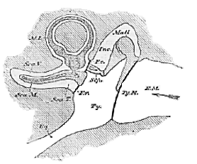
Fig. 70.–A Diagram illustrative of the relative Positions of the various Parts of the Ear.
E.M. external auditory meatus; Ty. M. tympanic membrane; Ty. tympanum; Mall.. malleus; Inc. incus; Stp. stapes; F.o. fenestra ovalis; F. r. fenestra rotunda; Eu. Eustachian tube; M.L. membranous labyrinth, only one semicircular canal with its ampulla being represented; Sca.V., Sca.T., Sca.M., the scalæ of the cochlea, which is supposed to be unrolled.
The rounded surface of the head of the malleus fits into a corresponding pit in the end of a third bone, the incus or anvil bone, which has two processes one, horizontal, which rests upon a support afforded to it by the walls of the tympanum; while the other, vertical, descends almost parallel with the long process of the malleus, and articulates with the stapes, or rather unites with a little bone, [208] the os orbiculare, which articulates with the stapes (Figs. 69 and 70).
The three bones thus form a chain between the fenestra ovalis and the tympanic membrane, and the whole series turns upon a horizontal axis, the two ends of which, formed by the horizontal process of the incus and the slender process of the malleus, rest in the walls of the tympanum. The general direction of this axis is represented by the line a b in Fig. 69, or by a line perpendicular to the plane of the paper, passing through the head of the malleus in Fig. 70. It follows, therefore, that whatever causes the membrane of the drum to vibrate backwards and forwards, must force the handle of the malleus to travel in the same way. This must cause a corresponding motion of the long process of the incus, the end of which must drag the stapes backwards and forwards. And, as this is fastened to the membrane of the fenestra ovalis which is in contact with the perilymph, it must set this fluid vibrating throughout its whole extent, the thrustings in of the membrane of the fenestra ovalis being compensated by corresponding thrustings out of the membrane of the fenestra rotunda, and vice versa.
The vibrations of the perilymph thus produced will affect the endolymph, and this the otolithes, hairs, or fibres; by which, finally, the auditory nerves will be excited.
21. The membrane of the fenestra ovalis and the tympanic membrane will necessarily vibrate the more freely the looser they are, and the reverse. But there are two muscles–one, called the stapedius, which passes from the floor of the tympanum to the orbicular bone, and the other, the tensor tympani, from the front wall of the drum to the malleus. Each of the muscles when it contracts tightens the membranes in question, and restricts their vibrations or, in other words, tends to check the effect of any cause which sets these membranes vibrating.
22. The outer extremity of the external meatus is surrounded by the concha or external ear (Co. Fig. 68), a broad, peculiarly-shaped, and for the most part cartilaginous plate, the general plane of which is at right angles with that of the axis of the auditory opening. The concha can be moved by most animals and by some human beings [209] in various directions by means of muscles, which pass to it from the side of the head.
23. The manner in which the complex apparatus now described intermediates between the physical agent, which is the condition of the sensation of sound, and the nervous expansion, the affection of which alone can excite that sensation, must next be considered.
All bodies which produce sound are in a state of vibration, and they communicate the vibrations of their own substance to the air with which they are in contact, and thus throw that air into waves, just as a stick waved backwards and forwards in water throws the water into waves.
The aerial waves, produced by the vibrations of sonorous bodies, in part enter the external auditory passage, and in part strike upon the concha of the external ear and the outer surface of the head. It may be that some of the latter impulses are transmitted through the solid structure of the skull to the organ of hearing; but before they reach it they must, under ordinary circumstances, have become so scanty and weak, that they may be left out of consideration.
The aerial waves which enter the meatus all impinge upon the membrane of the drum and set it vibrating, stretched membranes taking up vibrations from the air with great readiness.
24. The vibrations thus set up in the membrane of the tympanum are communicated, in part, to the air contained in the drum of the ear, and, in part, to the malleus, and thence to the other auditory ossicles.
The vibrations communicated to the air of the drum impinge upon the inner wall of the tympanum, on the greater part of which, from its density, they can produce very little effect. Where this wall is formed by the membrane of the fenestra rotunda, however, the communication of motion must necessarily be greater.
The vibrations which are communicated to the malleus and the chain of ossicles may be of two kinds: vibrations of the particles of the bones, and vibrations of the bones as a whole. If a beam of wood, freely suspended, be very gently scratched with a pin, its particles will be thrown into a state of vibration, as will be evidenced by the sound [210] given out, but the beam itself will not be moved. Again, if a strong wind blow against the beam, it will swing visibly, without any vibrations of its particles among themselves. On the other hand, if the beam be sharply struck with a hammer, it will not only give out a sound, showing that its particles are vibrating, but it will also swing from the impulse given to its whole mass.
Under the last-mentioned circumstances, a blind man standing near the beam would be conscious of nothing but the sound, the product of molecular vibration, or invisible oscillation of the particles of the beam; while a deaf man in the same position, would be aware of nothing but the visible oscillation of the beam as a whole.
25. Thus, to return to the chain of auditory ossicles, while it seems hardly to be doubted that, when the membrane of the drum vibrates, they may be set vibrating both as a whole and in their particles, it depends upon subsidiary arrangements whether the large vibrations, or the minute ones, shall make themselves obvious to the auditory nerve, which is in the position of our deaf, or blind, man.
The evidence at present is in favour of the conclusion, that it is the vibrations of the bones, as a whole, which are the chief agents in transmitting the impulses of the aerial waves.
For, in the first place, the disposition of the bones and the mode of their articulation are very much against the transmission of molecular vibrations through their substance, while, on the other hand, they are extremely favourable to their vibration en masse. The long processes of the malleus and incus swing, like a pendulum, upon the axis furnished by the short processes of these bones; while the mode of connection of the incus with the stapes, and of the latter with the edges of the fenestra ovalis, allows that bone free play, inwards and outwards. In the second place the total length of the chain of ossicles is very small compared with the length of the waves of audible sounds, and physical considerations teach us that in a like small rod, similarly capable of swinging en masse, the minute molecular vibrations would be inappreciable. Thirdly, it is affirmed, as the result of experiments, that the bone called columella, which, in birds, takes the place of the [211] chain of ossicles in man, does actually vibrate as a whole, and at the same rate as the membrane of the drum, when aerial vibrations strike upon the latter.
26. Thus, there is reason to believe that when the tympanic membrane is set vibrating, it causes the process of the malleus, which is fixed to it, to swing at the same rate; the head of the malleus consequently turns through a small arc on its pivot, the slender process. But the turning of the head of the malleus involves that of the head of the incus upon its pivot, the short process. In consequence the long process of the incus swings through an arc which has been estimated as being equal to about two-thirds of that described by the handle of the malleus. The extent of the push is thereby somewhat diminished, but the force of the push is proportionately increased, in so confined a space this change is advantageous. The long process, however, is so fixed to the stapes that it cannot vibrate without, to a corresponding extent and at the same rate, pulling this out of, and pushing it into, the fenestra ovalis. But every pull and push imparts a corresponding set of shakes to the perilymph, which fills the bony labyrinth and cochlea, external to the membranous labyrinth and scala media. These shakes are communicated to the endolymph and fluid of the scala media, and, by the help of the otolithes and the fibres of Corti, are finally converted into impulses, which act as irritants of the ends of the vestibular and cochlear divisions of the auditory nerve.
27. The difference between the functions of the membranous labyrinth (to which the vestibular nerve is distributed) and those of the cochlea are not quite certainly made out, but the following views have been suggested:–
The membranous labyrinth may be regarded as an apparatus whereby sounds are appreciated and distinguished according to their intensity or quantity; but which does not afford any means of discriminating their qualities. The vestibular nerve tells us that sounds are weak or loud, but gives us no impression of tone, or melody, or harmony.
The cochlea, on the other hand, it is supposed, enables the mind to discriminate the quality rather than the quantity or intensity of sound. It is suggested that the excitement of any single filament of the cochlear nerve [212] gives rise, in the mind, to a distinct musical impression; and that every fraction of a tone which a well-trained ear is capable of distinguishing is represented by its separate nerve-fibre. Under this view the scala media resembles a key-board, in function, as well as in appearance, the fibres of Corti being the keys, and the ends of the nerves representing the strings which the keys strike. If it were possible to irritate each of these nerve-fibres experimentally, we should be able to produce any musical tone, at will, in the sensorium of the person experimented upon, just as any note on a piano is produced by striking the appropriate key.
28. A tuning-fork may be set vibrating, if its own particular note, or one harmonic with it, be sounded in its neighbourhood. In other words, it will vibrate under the influence of a particular set of vibrations, and no others. If the vibrating ends of the tuning-fork were so arranged as to impinge upon a nerve, their repeated minute blows would at once excite this nerve.
Suppose that of a set of tuning-forks, tuned to every note and distinguishing fractions of a note in the scale, one were thus connected with the end of every fibre of the cochlear nerve; then any vibration communicated to the perilymph would affect the tuning-fork which could vibrate with it, while the rest would be absolutely, or relatively, indifferent to that vibration. In other words, the vibration would give rise to the sensation of one particular tone, and no other, and every musical interval would be represented by a distinct impression on the sensorium.
29. It is suggested that the fibres of Corti are competent to perform the function of such tuning-forks; that each of them is set vibrating to its full strength by a particular kind of wave sent through the perilymph, and by no other; and that each affects a particular fibre of the cochlear nerve only. But it must be remembered that the view here given is a suggestion only which, however probable, has not yet been proved. Indeed recent inquiries have rather diminished than increased its probability.
The fibres of the cochlear nerve may be excited by internal causes, such as the varying pressure of the blood and the like: and in some persons such internal influences do give rise to veritable musical spectra, sometimes of a [213] very intense character. But, for the appreciation of music produced external to us, we depend upon the intermediation of the scala media and its Cortian fibres.
30. It has already been explained that the stapedius and tensor tympani muscles are competent to tighten the membrane of the fenestra ovalis and that of the tympanum, and it is probable that they come into action when the sonorous impulses are too violent, and would produce too extensive vibrations of these membranes. They therefore tend to moderate the effect of intense sound, in much the same way that, as we shall find, the contraction of the circular fibres of the iris tends to moderate the effect of intense light in the eye.
The function of the Eustachian tube is, probably, to keep the air in the tympanum, or on the inner side of the tympanic membrane, of about the same tension as that on the outer side, which could not always be the case if the tympanum were a closed cavity.
1 I employ this term as the equivalent of canalis cochlearis. The true nature and connections of these parts have only recently been properly worked out, and the account now given will be found to be somewhat different from that in the first edition of this work, See particularly the explanation of Fig. 67.
[236]
The coalescence of sensations with one another and with other states of consciousness.
1. In explaining the functions of the sensory organs, I have hitherto confined myself to describing the means by which the physical agent of a sensation is enabled to irritate a given sensory nerve; and to giving some account of the simple sensations which are thus evolved.
Simple sensations of this kind are such as might be produced by the irritation of a single nerve-fibre, or of several nerve-fibres by the same agent. Such are the sensations of contact, of warmth, of sweetness, of an odour, of a musical note, of whiteness, or redness.
But very few of our sensations are thus simple. Most of even those which we are in the habit of regarding as simple, are really compounds of different sensations, or of sensations with ideas, or with judgments. For example, in the preceding cases, it is very difficult to separate the sensation of contact from the judgment that something is touching us; of sweetness, from the idea of something in the mouth; of sound or light, from the judgment that something outside us is shining, or sounding.
2. The sensations of smell are those which are least complicated by accessories of this sort. Thus, particles of musk diffuse themselves with great rapidity through the nasal passages, and give rise to the sensation of a powerful odour. But beyond a broad notion that the odour is in the nose, this sensation is unaccompanied by any ideas of locality and direction. Still less does it give rise to any conception of form, or size, or force, [237] or of succession, or contemporaneity. If a man had no other sense than that of smell, and musk were the only odorous body, he could have no sense of outness–no power of distinguishing between the external world and himself.
3. Contrast this with what may seem to be the equally simple sensation obtained by drawing the finger along the table, the eyes being shut. This act gives one the sensation of a flat, hard surface outside oneself, which appears to be just as simple as the odour of musk, but is really a complex state of feeling compounded of–
(a) Pure sensations of contact.
(b) Pure muscular sensations of two kinds,–the one arising from the resistance of the table, the other from the actions of those muscles which draw the finger along.
(c) Ideas of the order in which these pure sensations succeed one another.
(d) Comparisons of these sensations and their order, with the recollection of like sensations similarly arranged, which have been obtained on previous occasions.
(e) Recollections of the impressions of extension, flatness, &c. made on the organ of vision when these previous tactile and muscular sensations were obtained. Thus, in this case, the only pure sensations are those of contact and muscular action. The greater part of what we call the sensation is a complex mass of present and recollected ideas and judgments.
4. Should any doubt remain that we do thus mix up our sensations with our judgments into one indistinguishable whole, shut the eyes as before, and, instead of touching the table with the finger, take a round lead pencil between the fingers, and draw that along the table. The "sensation" of a flat hard surface will be just as clear as before; and yet all that we touch is the round surface of the pencil, and the only pure sensations we owe to the table are those afforded by the muscular sense. In fact, in this case, our "sensation" of a flat hard surface is entirely a judgment based upon what the muscular sense tells us is going on in certain muscles.
A still more striking case of the tenacity with which we adhere to complex judgments, which we conceive to be pure sensations, and are unable to analyse otherwise [238] than by a process of abstract reasoning, is afforded by our sense of roundness.
Anyone taking a marble between two fingers will say that he feels it to be a single round body; and he will probably be as much at a loss to answer the question how he knows that it is round, as he would be if he were asked how he knows that a scent is a scent.
Nevertheless, this notion of the roundness of the marble is really a very complex judgment, and that it is so may be shown by a simple experiment. If the index and middle fingers be crossed, and the marble placed between them, so as to be in contact with both, it is utterly impossible to avoid the belief that there are two marbles instead of one. Even looking at the marble, and seeing that there is only one, does not weaken the apparent proof derived from touch that there are two.1
The fact is, that our notions of singleness and roundness are, really, highly complex judgments based upon a few simple sensations; and when the ordinary conditions of those judgments are reversed, the judgment is also reversed.
With the index and the middle fingers in their ordinary position, it is of course impossible that the outer sides of each should touch opposite surfaces of one spheroidal body. If, in the natural and usual position of the fingers, their outer surfaces simultaneously give us the impression of a spheroid (which itself is a complex judgment), it is in the nature of things that there must be two spheroids. But, when the fingers are crossed over the marble, the outer side of each finger is really in contact with a spheroid; and the mind, taking no cognizance of the crossing, judges in accordance with its universal experience, that two spheroids, and not one, give rise to the sensations which are perceived
5. Phenomena of this kind are not uncommonly called delusions of the senses; but there is no such thing as a fictitious, or delusive, sensation. A sensation must [239] exist to be a sensation, and, if it exists, it is real and not delusive. But the judgments we form respecting the causes and conditions of the sensations of which we are aware, are very often erroneous and delusive enough; and such judgments may be brought about in the domain of every sense, either by artificial combinations of sensations, or by the influence of unusual conditions of the body itself. The latter give rise to what are called subjective sensations.
Mankind would be subject to fewer delusions than they are, if they constantly bore in mind their liability to false judgments due to unusual combinations, either artificial or natural, of true sensations. Men say, "I felt," "I heard," "I saw" such and such a thing, when, in ninety-nine cases out of a hundred, what they really mean is, that they judge that certain sensations of touch, hearing, or sight, of which they were conscious, were caused by such and such things.
6. Among subjective sensations within the domain of touch, are the feelings of creeping and prickling of the skin, which are not uncommon in certain states of the circulation. The subjective evil smells and bad tastes which accompany some diseases are very probably due to similar disturbances in the circulation of the sensory organs of smell and taste.
Many persons are liable to what may be called auditory spectra–music of various degrees of complexity sounding in their ears, without any external cause, while they are wide awake. I know not if other persons are similarly troubled, but in reading books written by persons with whom I am acquainted, I am sometimes tormented by hearing the words pronounced in the exact way in which these persons would utter them, any trick or peculiarity of voice, or gesture, being, also, very accurately reproduced. And I suppose that everyone must have been startled, at times, by the extreme distinctness with which his thoughts have embodied themselves in apparent voices.
The most wonderful exemplifications of subjective sensation, however, are afforded by the organ of sight.
Anyone who has witnessed the sufferings of a man labouring under delirium tremens (a disease produced by excessive drinking), from the marvellous distinctness of [240] his visions, which sometimes take the forms of devils, sometimes of creeping animals, but almost always of something fearful or loathsome, will not doubt the intensity of subjective sensations in the domain of vision.
7. But that illusive visions of great distinctness should appear, it is not necessary for the nervous system to be thus obviously deranged. People in the full possession of their faculties, and of high intelligence, may be subject to such appearances, for which no distinct cause can be assigned. An excellent illustration of this is the famous case of Mrs. A. given by Sir David Brewster, in his "Natural Magic." (See Appendix.)
It should be mentioned that Mrs. A. was naturally a person of very vivid imagination, and that, at the time the most notable of these illusions appeared, her health was weak from bronchitis and enfeebled digestion.
It is obvious that nothing but the singular courage and clear intellect of Mrs. A. prevented her from becoming a mine of ghost stories of the most excellently authenticated kind. And the particular value of her history lies in its showing, that the clearest testimony of the most unimpeachable witness may be quite inconclusive as to the objective reality of something which the winless has seen.
Mrs. A. undoubtedly saw what she said she saw. The evidence of her eyes as to the existence of the apparitions, and of her ears to those of the voices, was, in itself, as perfectly trustworthy as their evidence would have been had the objects really existed. For there can be no doubt that exactly those parts of her retina which would have been affected by the image of a cat, and those parts of her auditory organ which would have been set vibrating by her husband's voice, or the portions of the sensorium with which those organs of sense are connected, were thrown into a corresponding state of activity by some internal cause.
What the senses testify is neither more nor less than the fact of their own affection. As to the cause of that affection they really say nothing, but leave the mind to form its own judgment on the matter. A hasty or superstitious person in Mrs. A.'s place would have formed a wrong judgment, and would have stood by it on the plea that "she must believe her senses."
[241] 8. The delusions of the judgment, produced not by abnormal conditions of the body, but by unusual or artificial combinations of sensations, or by suggestions of ideas, are exceedingly numerous, and, occasionally are not a little remarkable.
Some of those which arise out of the sensation of touch have already been noted. I do not know of any produced through smell or taste, but hearing is a fertile source of such errors.
What is called ventriloquism (speaking from the belly), and is not uncommonly ascribed to a mysterious power of producing voice somewhere else than in the larynx, depends entirely upon the accuracy with which the performer can simulate sounds of a particular character, and upon the skill with which he can suggest a belief in the existence of the causes of these sounds. Thus, if the ventriloquist desire to create the belief that a voice issues from the bowels of the earth, he imitates with great accuracy the tones of such a half-stifled voice, and suggests the existence of some one uttering it by directing his answers and gestures towards the ground. These gestures and tones are such as would be produced by a given cause; and no other cause being apparent, the mind of the bystander insensibly judges the suggested cause to exist.
9. The delusions of the judgment through the sense of sight–optical delusions, as they are called–are more numerous than any others, because such a great number of what we think to be simple visual sensations are really very complex aggregates of visual sensations, tactile sensations, judgments, and recollections of former sensations and judgments.
It will be instructive to analyse some of these judgments into their principles, and to explain the delusions by the application of these principles.
10. When an external body is felt by the touch to be in a given place, the image of that body falls on a point of the retina which lies at one end of a straight tine joining the body and the retina, and traversing a particular region of the centre of the eye. This straight line is called the Optic Axis.
Conversely, when any part of the surface of the retina [242] is excited, the luminous sensation is referred by the mind to some point outside the body, in the direction of the optic axis
It is for this reason that when a phosphene is created by pressure, say on the outer and lower side of the eyeball, the luminous image appears to lie above, and to the inner side of, the eye. Any external object which could produce the sense of light in the part of the retina pressed upon must, owing to the inversion of the retinal images (see Lesson IX. § 23), in fact occupy this position; and hence the mind refers the light seen to an object in that position.
11. The same kind of explanation is applicable to the apparent paradox that, while all the pictures of external objects are certainly inverted on the retina by the refracting media of the eye, we nevertheless see them upright. It is difficult to understand this, until one reflects that the retina has, in itself, no means of indicating to the mind which of its parts lies at the top, and which at the bottom, and that the mind learns to call an impression on the retina high or low, right or left, simply on account of the association of such an impression with certain coincident tactile impressions. In other words, when one part of the retina is affected, the object causing the affection is found to be near the right hand; when another, the left; when another, the hand has to be raised to reach the object; when yet another, it has to be depressed to reach it. And thus the several impressions on the retina are called right, left, upper, lower, quite irrespectively of their real positions, of which the mind has, and can have, no cognizance.
12. When an external body is ascertained by touch to be simple, it forms but one image on the retina of a single eye; and when two or more images fall on the retina of a simple eye, they ordinarily proceed from a corresponding number of bodies which are distinct to the touch.
Conversely, the sensation of two or mare images is judged by the mind to proceed from two or more objects.
If two pin-holes be made in a piece of cardboard at a distance less than the diameter of the pupil, and a small object like the head of a pin be held pretty close to the eye, and viewed through these holes, two images of the [243] head of the pin will be seen, The reason of this is, that the rays of light from the head of the pin are split by the card into two minute pencils, which pass into the eye on either side of its centre, and cannot be united again and brought to one focus on account of the nearness of the pin to the eye. Hence they fall on different parts of the retina, and each pencil of rays, being very small, makes a tolerably distinct image of its own of the pin's head on the retina. Each of these images is now referred outward (§ 10) in the direction of the appropriate optic axis, and two pins are apparently seen instead of one. A like explanation applies to multiplying glasses and doubly refracting crystals, both of which, in their own ways, split the pencils of light proceeding from a single object into two or more separate bundles. These give rise to as many images, each of which is referred by the mind to a distinct external object.
13. Certain visual phenomena ordinarily accompany those products of tactile sensation to which we give the name of size, distance, and form. That, other thing being alike, the space of the retina covered by the image of a large object is larger than that covered by a small object; while that covered by a near object is larger than that covered by a [distant] object; and, other conditions being alike, a near object is more brilliant than a distant one. Furthermore, the shadows of objects differ with the forms of their surfaces, as determined by touch.
Conversely, if these visual sensations can he produced, they inevitably suggest a belief in the existence of objects competent to produce the corresponding tactile sensations.
What is called perspective, whether solid or aërial, in drawing, or painting, depends on the application of these principles. It is a kind of visual ventriloquism–the painter putting upon his canvas all the conditions requisite for the production of images on the retina, having the size, relative form, and intensity of colour of those which would actually be produced by the objects themselves in nature. And the success of his picture, as an imitation, depends upon the closeness of the resemblance between the images it produces on the retina, and those which would be produced by the objects represented.
To most persons the image of a pin, at five or six [244] inches from the eye, appears blurred and indistinct–the eye not being capable of adjustment to so short a focus. If a small hole be made in a piece of card, the circumferential rays which cause the blur are cut off, and the image becomes distinct. But at the same time it is magnified, or looks bigger, because the image of the pin, in spite of the loss of the circumferential rays, occupies a much larger extent of the retina when close than when distant. All convex glasses produce the same effect–while concave lenses diminish the apparent size of an object, because they diminish the size of its image on the retina.
15. The moon, or the sun, when near the horizon appear very much larger than they are when high in the sky. When in the latter position, in fact, we have nothing to compare them with, and the small extent of the retina which their images occupy suggests small absolute size. But as they set, we see them passing behind great trees and buildings which we know to be very large and very distant, and yet occupying a larger space on the retina than the latter do. Hence the vague suggestion of their larger size.
16. If a convex surface be lighted from one side, the side towards the light is bright–that turned from the light, dark or in shadow; while a concavity is shaded on the side towards the light, bright on the opposite side.
If a new half-crown, or a medal with a well-raised head upon its face, be lighted sideways by a candle, we at once know the head to be raised (or a cameo) by the disposition of the light and shade; and if an intaglio, or medal on which the head is hollowed out, be lighted in the same way, its nature is as readily judged by the eye.
But now, if either of the objects thus lighted be viewed with a convex lens, which inverts its position, the light and dark sides will be reversed. With the reversal the judgment of the mind will change, so that the cameo will be regarded as an intaglio, and the intaglio as a cameo; for the light still comes from where it did, but the cameo appears to have the shadows of an intaglio, and vice versa. So completely, however, is this interpretation of the facts as a matter of judgment, that if a pin be stuck beside the medal so as to throw a shadow, the pin and its shadows being reversed by the lens, will suggest that the direction [245] of the light is also reversed, and the medals will seem to be what they really are.
17. Whenever an external object is watched rapidly changing its form, a continuous series of different pictures of the object is impressed upon the same spot of the retina.
Conversely, if a continuous series of different pictures of one object is impressed upon one part of the retina, the mind judges that they are due to a single external object, undergoing changes of form.
This is the principle of the curious toy called the thaumatrope, or "zootrope," or "wheel of life," by the help of which, on looking through a hole, one sees images of jugglers throwing up and catching balls, or boys playing at leapfrog over one another's backs. This is managed by painting at intervals, on a disk of card, figures and jugglers in the attitudes of throwing, waiting to catch, and catching; or boys "giving a back," leaping, and coming into position after leaping. The disk is then made to rotate before an opening, so that each image shall be presented for an instant, and follow its predecessor before the impression of the latter has died away. The result is that the succession of different pictures irresistibly suggests one or more objects undergoing successive changes–the juggler seems to throw the balls, and the boys appear to jump over one another's backs.
18. When an external object is ascertained by touch to be single, the centres of its retinal images in the two eyes fall upon the centres of the yellow spots of the two eyes, when both eyes are directed towards it; but if there be two external objects, the centres of both their images cannot fall at the same time, upon the centres of the yellow spots.
Conversely, when the centres of two images, formed simultaneously in the two eyes, fall upon the centres of the yellow spots, the mind judges the images to be caused by a single external object; but if not, by two.
This seems to be the only admissible explanation of the facts, that an object which appears single to the touch and when viewed with one eye, also appears single when it is viewed with both eyes, though two images of it are necessarily formed; and on the other hand, that when the centres of the two images of one object do not fall on the [246] centres of the yellow spots, both images are seen separately, and we have double vision. In squinting, the axes of the two eyes do not converge equally towards the object viewed. In consequence of this, when the centre of the image formed by one eye falls on the centre of the yellow spot, the corresponding part of that formed by the other eye does not, and double vision is the result.
For simplicity's sake we have supposed the images to fall on the centre of the yellow spot. But though vision is distinct only in the yellow spot, it is not absolutely limited to it; and it is quite possible for an object to be seen as a single object with two eyes, though its images fall on the two retinas outside the yellow spots. All that is necessary is that the two spots of the retinas on which the images fall should be similarly disposed towards the centres of their respective yellow spots. Any two points of the two retinas thus similarly disposed towards their respective yellow spots (or more exactly to the points in which the optic axes end), are spoken of as corresponding points; and any two images covering two corresponding areas are conceived of as coming from a single object. It is obvious that the inner (or nasal) side of one retina corresponds to the outer (or cheek) side of the other.
19. In single vision with two eyes, the axes of the two eyes, of the movements of which the muscular sense gives an indication, cut one another at a greater angle when the object approaches, at a less angle when it goes further off.
Conversely, if without changing the position of an object, the axes of the two eyes which view it can be made to converge or diverge, the object will seem to approach or go further off.
In the instrument called the pseudoscope, mirrors or prisms are disposed in such a manner that the angle at which rays of light from an object enter the two eyes, can be altered without any change in the object itself; and consequently the axes of these eyes are made to converge or diverge. In the former case the object seems to approach; in the latter, to recede.
20. When a body of moderate size, ascertained by touch to be solid, is viewed with both eyes, the images of it, formed by the two eyes, are necessarily different (one showing more of its right side, the other of its left side). [247] Nevertheless, they coalesce into a common image, which gives the impression of solidity.
Conversely, if the two images of the right and left aspects of a solid body be made to fall upon the retinas of the two eyes in such a way as to coalesce into a common image, they are judged by the mind to proceed from the single solid body which alone, under ordinary circumstances, is competent to produce them.
The stereoscope is constructed upon this principle. Whatever its form, it is so contrived as to throw the images of two pictures of a solid body, such as would be obtained by the right and left eye of a spectator, on to such parts of the retinas of the person who uses the stereoscope as would receive these images, if they really proceeded from one solid body. The mind immediately judges them to arise from a single external solid body, and sees such a solid body in place of the two pictures.
The operation of the mind upon the sensations presented to it by the two eyes is exactly comparable to that which takes place when, on holding a marble between the finger and thumb, we at once declare it to be a single sphere (§ 4). That which is absolutely presented to the mind by the sense of touch in this case is by no means the sensation of one spheroidal body, but two distinct sensations of two convex surfaces. That these two distinct convexities belong to one sphere, is an act of judgment, or process of unconscious reasoning, based upon many particulars of past and present experience, of which we have, at the moment, no distinct consciousness
1 A ludicrous form of this experiment is to apply the crossed fingers to the end of the nose, when it at once appears double; and, in spite of the absurdity of the conviction, the mind cannot expel it, so long as the sensations last.
[297]
A Table of Anatomical and Physiological Constants
The weight of the body of a full-grown man may be taken at 154 lbs.
I. GENERAL STATISTICS
| Such a body would be made up of | lbs. | |
| Muscles and their appurtenances | 68 | |
| Skeleton | 24 | |
| Skin | 10-1/2 | |
| Fat | 28 | |
| Brain | 3 | |
| Thoracic viscera | 2-1/2 | |
| Abdominal viscera | 11 | |
| 1471 | ||
| Or of | lbs. | |
| Water | 88 | |
| Solid matters | 66 | |
[298] The solids would consist of the elements oxygen, hydrogen, carbon, nitrogen, phosphorus, sulphur, silicon, chlorine, fluorine, potassium, sodium, calcium (lithium), magnesium, iron (manganese copper), lead, and may be arranged under the heads of–
Proteids. Amyloids. Fats. Minerals.
Such a body would lose in 24 hours–of water, about 40,000 grains, or 6 lbs.; of other matters about 14,500 grains, or over 2 lbs.; among which of carbon 4,000 grains; of nitrogen 300 grains; of mineral matters 400 grains; and would part, per diem, with as much heat as would raise 8,700 lbs. of water 0° to 1° Fahr., which is equivalent to 3,000 foot-tons.2
The losses would occur through various organs, thus–by
| Water grs. |
Other Matter grs. |
N. grs. |
C. grs. | |
| Lungs | 5,000 | 12,000 | ... | 3,300 |
| Kidneys | 23,000 | 1,000 | 250 | 140 |
| Skin | 10,000 | 700 | 10 | 100 |
| Fæces | 2,000 | 800 | 40 | 460 |
| Total | 40,000 | 14,500 | 300 | 4,000 |
The gains and losses of the body would be as follows:–
| grs. | ||
| Creditor | Solid dry food | 8,000 |
| Oxygen | 10,000 | |
| Water | 36,500 | |
| 54,500 | ||
| grs. | ||
| Debtor | Water | 40,000 |
| Other Matters | 14,500 | |
| 54,500 |
[299]
Such a body would require for daily food, carbon 4,000 grains, nitrogen, 300 grains; which, with the other necessary elements, would be most conveniently disposed in– which, in turn, might be obtained, for instance, by means of– The fæces passed, per diem, would amount to about 2,800 grains, containing solid matter 800 grains. III. CIRCULATION
In such a body the heart would beat 75 times a minute, and probably drive out, at each stroke from each ventricle, from 5 to 6 cubic inches, or about l,500 grains of blood. The blood would probably move in the great arteries at a rate of about 12 inches in a second, in the capillaries at 1 to 1-1/2 inches in a minute; and the time taken up in performing the entire circuit would probably be about 30 seconds. The left ventricle would probably exert a pressure on the aorta equal to the pressure on the square-inch of a column of blood about 9 feet in height; or of a column of [300] mercury about 9-1/2 inches in height; and would do in 24 hours an amount of work equivalent to about 90 foot-tons; the work of the whole heart being about 120 foot-tons.
IV. RESPIRATION
Such a body would breathe 15 times a minute. The lungs would contain of residual air about 100 cubic inches, of supplemental or reserve air about 100 cubic inches, of tidal air 20 to 30 cubic inches, and of complemental air 100 cubic inches. The vital capacity of the chest–that is, the greatest quantity of air which could be inspired or expired–would be about 230 cubic inches. There would pass through the lungs, per diem, about 350 cubic feet of air. In passing through the lungs, the air would lose from 4 to 6 per cent. of its volume of oxygen, and gain 4 to 5 per cent. of carbonic acid. During 24 hours there would be consumed about 10,000 grains oxygen; and produced about 12,000 grains carbonic acid, corresponding to 3,300 grains carbon. During the same time about 5,000 grains or 9 oz. of water would be exhaled by the lungs. In 24 hours such a body would vitiate 1750 cubic feet of pure air to the extent of 1 per cent., or 17,500 cubic feet of pure air to the extent of 1 per 1,000. Taking the amount of carbonic acid in the atmosphere at 3 parts, and in expired air at 470 parts in 10,000, such a body would require a supply per diem of more than 23,000 cubic feet of ordinary air, in order that the surrounding atmosphere might not contain more than 1 per l,000 of carbonic acid (when air is vitiated from animal sources with carbonic acid to more than 1 per l,000, the concomitant impurities become appreciable to the nose). A man of the weight mentioned (11 stone) ought, therefore, to have at least 800 cubic feet of well-ventilated space.
V. CUTANEOUS EXCRETION
Such a body would throw off by the skin–of water about 18 ounces, or 10,000 grains; of solid matters about 300 grains; of carbonic acid about 400 grains, in 24 hours.
[301]
Such a body would pass by the kidneys–of water about 50 ounces; of urea about 500 grains; of other solid matters about 500 grains, in 24 hours.
VII. NERVOUS ACTION
In the frog a nervous impulse travels at the rate of about 80 feet in a second. In a man a nervous (sensory) impulse has been variously calculated to travel at 100, 200, or 300 feet in a second.
VIII. HISTOLOGY.
Red corpuscles of the blood are about 1/3200th of an inch in breadth; white corpuscles 1/2500th. Striated muscular fibers are about 1/400th of an inch in breadth; plain 1/400th. Nerve-fibres vary between 1/1500 and 1/12000 of an inch in breadth. Connective tissue fibres are about 1/4000th of an inch in breadth. Epithelium scales (of the skin) are about 1/500th of an inch in breadth. Capillary blood-vessels are from 1/3500th to 1/2000 of an inch in breadth. Cilia (from the wind-pipe) are about 1/3000th of an inch in length.
The cones in the "yellow spot" of the retina are about 1/10000 of an inch in breadth.
1 The addition of 7 lbs. of blood, the quantity which will readily drain away from the body, will bring the total to 154 lbs. A considerable quantity of blood will, however, always remain in the capillaries and small blood-vessels, and must be reckoned with the various tissues. The total quantity of blood in the body is now calculated at about 1-13th of the body weight, i.e. , about 12 lbs. 2 A foot-ton is the equivalent of the work required to lift one ton one foot high.
Appendix B. The Case of Mrs. A––. (See p. 240.)
[302] (1) The first illusion to which Mrs. A. was subject, was one which affected only the ear. On the 21st of December, 1830, about half-past four in the afternoon, she was standing near the fire in the hall, and on the point of going up to dress, when she heard, as she supposed, her husband's voice calling her by name: "––,––, come here. Come to me!" She imagined that he was calling at the door to have it opened; but upon going there and opening the door, she was surprised to find no person there. Upon returning to the fire she again heard the same voice calling out very distinctly and loudly, "––, come, come here!" She then opened two other doors of the same room, and upon seeing no person, she returned to the fireplace. After a few moments she heard the same voice calling, "Come to me, come! come away!" in a loud, plaintive, and somewhat impatient tone; she answered as loudly, "Where are you? I don't know where you are," still imagining that he was somewhere in search of her; upon receiving no answer, she shortly went upstairs. On Mr. A.'s return to the house, about half an hour afterwards, she inquired why he had called her so often he was, and where he. and she was of course greatly surprised to learn that he had not been near the house at the time. A similar illusion, which excited no particular notice at the time, occurred to Mrs. A. when residing at Florence, about ten years before, and when she was in perfect health. When she was undressing after a ball, she heard a voice call her repeatedly by name, and she was at that time unable to account for it. [303] (2) The next illusion which occurred to Mrs. A. was of a more alarming character. On the 30th of December, about four o'clock in the afternoon, Mrs. A. came downstairs into the drawing-room, which she had quitted only a few minutes before, and, on entering the room, she saw her husband, as she supposed, standing with his back to the fire. As he had gone out to take a walk about half an hour before, she was surprised to see him there, and asked him why he had returned so soon. The figure looked fixedly at her with a serious and thoughtful expression of countenance, but did not speak. Supposing that his mind was absorbed in thought, she sat down in an arm-chair near the fire, and within two feet, at most, of the figure, which she still saw standing before her. As its eyes, however, still continued to be fixed upon her, she said, after the lapse of a few minutes, "Why don't you speak?" The figure immediately moved off towards the window at the further end of the room, with its eyes still gazing on her, and it passed so very close to her in doing so, that she was struck with the circumstance of hearing no step or sound, nor feeling her clothes brushed against, nor even any agitation in the air. Although she was now convinced that the figure was not her husband, yet she never for a moment supposed that it was anything supernatural, and was soon convinced that it was a spectral illusion. As soon as this conviction had established itself in her mind, she recollected the experiment which I had suggested of trying to double the object; but before she was able distinctly to do this, the figure had retreated to the window, where it disappeared. Mrs. A. immediately followed it, shook the curtains, and examined the window, the impression having been so distinct and forcible that she was unwilling to believe that it was not a reality. Finding, however, that the figure had no natural means of escape, she was convinced that she had seen a spectral apparition like that recorded in Dr. Hibbert's work, and she consequently felt no alarm or agitation. The appearance was seen in bright daylight and lasted four or five minutes. When the figure stood close to her, it concealed the real objects behind it, and he apparition was fully as vivid as the reality (3) On these two occasions Mrs. A. was alone, but when [304] the next phantom appeared, her husband was present. This took place on the 4th of January, 1830. About ten o'clock at night, when Mr. and Mrs. A. were sitting in the drawing-room, Mr. A. took up the poker to stir the fire, and when he was in the act of doing this, Mrs. A. exclaimed, "Why, there's the cat in the room!" "Where?" exclaimed Mr. A. "There, close to you," she replied. "Where?" he repeated. "Why, on the rug, to be sure, between yourself and the coal-scuttle." Mr. A., who still had the poker in his hand, pushed it in the direction mentioned. "Take care," cried Mrs. A., "take care! you are hitting her with the poker." Mr. A. again asked her to point out exactly where she saw the cat. She replied, "Why, sitting up there close to your feet on the rug; she is looking at me. It is Kitty–come here, Kitty!" There were two cats in the house, one of which went by this name, and they were rarely, if ever, in the drawing-room. At this time Mrs. A. had no idea that the sight of the cat was an illusion. When she was asked to touch it, she got up for the purpose, and seemed as if she was pursuing something which moved away. She followed a few steps, and then said, "It has gone under the chair." Mr. A. assured her that it was an illusion, but she would not believe it. He then lifted up the chair, and Mrs. A. saw nothing more of it. The room was searched all over, and nothing found in it. There was a dog lying on the hearth, who would have betrayed great uneasiness if a cat had been in the room, but he lay perfectly quiet. In order to be quite certain, Mr. A. rang the bell, and sent for the cats, both of which were found in the housekeeper's room. (4) About a month after this occurrence, Mrs. A., who had taken a somewhat fatiguing drive during the day, was preparing to go to bed about eleven o'clock at night, and, sitting before the dressing-glass, was occupied in arranging her hair. She was in a listless and drowsy state of mind, but fully awake. When her fingers were in active motion among the papillotes, she was suddenly startled by seeing in the mirror the figure of a near relative, who was then in Scotland, and in perfect health. The apparition appeared over her left shoulder, and its eyes met hers in the glass. It was enveloped in grave-clothes, [305] closely pinned, as is usual with corpses, round the head and under the chin; and, though the eyes were open, the features were solemn and rigid. The dress was evidently a shroud, as Mrs. A. remarked even the punctured pattern usually worked in a peculiar manner round the edges of that garment. Mrs. A. described herself as, at the time, sensible of a feeling like what we conceive of fascination compelling her, for the time, to gaze upon this melancholy apparition, which was as distinct and vivid as any reflected reality could be, the light of the candle upon the dressing-table appearing to shine fully upon its face. After a few minutes she turned round to look for the reality of the form over her shoulder, but it was not visible, and it had also disappeared from the glass when she looked again in that direction.
* * * * * *
(7) On the 17th March, Mrs. A. was preparing for bed. She had dismissed her maid, and was sitting with her feet in hot water. Having an excellent memory, she had been thinking upon and repeating to herself a striking passage in the Edinburgh Review, when, on raising her eyes, she saw seated in a large easy-chair before her the figure of a deceased friend, the sister of Mr. A. The figure was dressed, as had been usual with her, with great neatness, but in a gown of a peculiar kind, such as Mrs. A. had never seen her wear, but exactly such as had been described to her by a common friend as having been worn by Mr. A.'s sister during her last visit to England. Mrs. A. paid particular attention to the dress, air, and appearance of the figure, which sat in an easy attitude in the chair, holding a handkerchief in one hand. Mrs. A. tried to speak to it, but experienced a difficulty in doing so, and in about three minutes the figure disappeared. About a minute afterwards, Mr. A. came into the room, and found Mrs. A. slightly nervous, but fully aware of the delusive nature of the apparition. She described it as having all the vivid colouring and apparent reality of life; and for some hours preceding this and other visions, she experienced a peculiar sensation in her eyes, which seemed to be relieved when the vision had ceased.
* * * * * *
[306] (9) On the 11th October, when sitting in the drawing-room, on one side of the fire-place, she saw the figure of another deceased friend moving towards her from the window at the farther end of the room. It approached the fire-place, and sat down in the chair opposite. As there were several persons in the room at the time, she describes the idea uppermost in her mind to have been a fear lest they should be alarmed at her staring, in the way she was conscious of doing, at vacancy, and should fancy her intellect disordered. Under the influence of this fear, and recollecting a story of a similar effect in your1 work on Demonology, which she had lately read, she summoned up the requisite resolution to enable her to cross the space before the fire-place, and seat herself in the same chair with the figure. The apparition remained perfectly distinct till she sat down, as it were, in its lap, when it vanished.
1 Sir Walter Scott: to whom Sir David Brewster's Letters on Natural Magic were addressed.
grs. Proteids 2,000 Amyloids 4,400 Fats 1,200 Minerals 400 Water 36,500 Total 44,500
grs. Lean beefsteaks 5,000 Bread 6,000 Milk 7,000 Potatoes 3,000 Butter, dripping, &c. 600 Water 22,900 Total 44,500
PREVIEW
TABLE of CONTENTS
BIBLIOGRAPHIES
1.
THH Publications
2.
Victorian Commentary
3.
20th Century Commentary
INDICES
1. Letter Index
2. Illustration Index
TIMELINEFAMILY TREE Gratitude and Permissions
C. Blinderman & D. Joyce
Clark University
1998
THE
HUXLEY
FILE
GUIDES § 1.
THH: His Mark § 2.
Voyage of the Rattlesnake § 3.
A Sort of Firm § 4.
Darwin's Bulldog § 5.
Hidden Bond: Evolution § 6.
Frankensteinosaurus § 7.
Bobbing Angels: Human Evolution § 8.
Matter of Life: Protoplasm § 9.
Medusa § 10.
Liberal Education § 11.
Scientific Education § 12.
Unity in Diversity § 13.
Agnosticism § 14.
New Reformation § 15.
Verbal Delusions: The Bible § 16.
Miltonic Hypothesis: Genesis § 17.
Extremely Wonderful Events: Resurrection and Demons § 18.
Emancipation: Gender and Race § 19.
Aryans et al.: Ethnology § 20.
The Good of Mankind § 21.
Jungle Versus Garden