 |
 |
| FRS
Fellow, Royal Society 1851 |
Nobel Swell
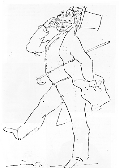
T. H. H. caricature of his having won R.S. gold medal |
Huxley Archives
 |
 |
| FRS
Fellow, Royal Society 1851 |
Nobel Swell
T. H. H. caricature of his having won R.S. gold medal |
Huxley Archives
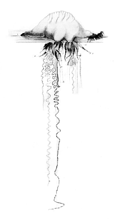
A Description of the Calycophoridæ and Physophoridæ observed during the voyage of H.M.S. "Ratttlesnake" in the years 1846-1850 (sel.) (1858) The Ray Society
[1] Sect. 1 MORPHOLOGY OF THE HYDROZOA.
The body of every Hydrozoon is essentially a sac, composed of two membranes, an external and an internal, which have been conveniently denominated by the terms ectoderm and endoderm . The cavity of the sac, which will be called the somatic cavity, contains a fluid, charged with nutritive matter in solution, and sometimes, if not always, with suspended solid particles, which performs the functions of the blood in animals of higher organization, and may be termed the somatic fluid . The ectoderm is commonly ciliated, at any rate while young; the endoderm is also very generally ciliated, though not always, nor in all parts. The cilia of the endoderm, aided by the contractions of the walls of the body, are the sole means provided by nature for the circulation of the nutritive fluid in the Hydrozoa; the cilia of the ectoderm, similarly aided by contractility, constitute the only respiratory mechanism.
Notwithstanding the extreme variety of form exhibited by the Hydrozoa, and the multiplicity and complexity of the organs which some of them possess, they never lose the traces of this primitive simplicity of organization; and it is but rarely that it is even disguised to any considerable extent. I know of no Hydrozoon, in which the two primary membranes, but little altered, cannot be at once detected in the walls of almost every part of the organism.
This important and obvious structural peculiarity could hardly escape notice, and I find it to have been observed by Trembley, Baker, Laurent, Corda, and Ecker, in Hydra ; by Rathke, in Coryne ; by Frey and Leuckart, in Lucernaria ; and it is given as a character of the Hydroid polypes in general (Hydra, Corynidæ, and Sertulariadæ) in the second edition of Cuvier's 'Leçons.' I pointed it out as the general law of structure of the Hydroid polypes, Diphydæ, and Physophoridæ, in a paper1 sent to the Linnean Society, from Australia, in 1847, but not read before that body until January, 1849; and I extended the generalization to the whole of the Hydrozoa in a 'Memoir on the Anatomy and Affinity of the Medusæ,' read before the Royal Society in June, 1849.
[2] Professor Allman, in his valuable 'Memoire on Cordylophora' ('Philo. Trans.,' 1853), has adopted and confirmed this morphological law, introducing the convenient terms "ectoderm and endoderm," to denote the inner and outer membranes; and Gegenbaur ('Beiträge zur näharen Kenntniss der Schwimmpolypen,' 1854, p. 42) has partially noticed its exemplification in Apolemia and Rhizophysa, but it seems, singularly enough, to have failed to attract the attention of the other excellent German observers, to whose late important investigations I shall so often have occasion to adver.
The peculiarity in the structure of the body-walls of the Hydrozoa, to which I have just referred, possesses a singular interest in its bearing upon the truth (for, with due limitation, it is a great truth) that there is a certain similarity between the adult stages of the lower animals and the embryonic conditions of those of higher organization.
For it is well known that, in a very early state, the germ, even of the highest animals, is a more or less complete sac, whose thin wall is divisible into two membranes, an inner and an outer; the latter, turned towards the external world; the former, in relation with the nutritive liquid–the yelk. The inner layer, as Remak has more particularly shown, undergoes but little histological change, and, throughout life, remains more particularly devoted to the function of alimentation, while the outer gives rise, by manifold differentiations of its tissue, to those complex structures which we know as integument, bones, muscles, nerves and sensory apparatus, and which especially subserve the functions of relation. At the same time the various organs are produced by a process of budding from one, or other, or both, of these primary layers of the germ.
Just so in the Hydrozoon: the ectoderm gives rise to the hard tegumentary tissues, to the more important masses of muscular fibre, and to those organs which we have every reason to believe are sensory, while the endoderm undergoes but very little modification. And every organ of a Hydrozoon is produced by budding from one, or other, or both, of these primitive membranes; the ordinary case being, that the new part commences its existence as a papillary process of both membranes, including, of course, a cæcal diverticulum of the somatic cavity.
Thus there is a very real and genuine analogy between the adult Hydrozoon and the embryonic vertebrate animal; but I need hardly say that it by no means justifies the assumption that the Hydrozoa are in any sense "arrested developments" of higher organisms. All that can justly be affirmed is, that the Hydrozoon travels for a certain distance along the same great highway of development as the higher animals, before it turns off to follow the road which leads to its special destination.
The entire double-walled body of the Hydrozoon, whether it be a minute, simple, oval sac, as in the embryonic condition, or such a vast and complex mass as a tree of Plumularia, and Agalma three feet long, or a Rhizostoma of still more massive proportions, will be termed, in the course of the ensuing pages, a hydrosoma .
The simplest condition of this hydrosoma is that observable in the common fresh-water Hydra, one end of whose body is expanded into a kind of disc, whereby the creature adheres to its support, while the opposite extremity presents a widely-open mouth, opening into a cavity which extends through the whole length of the animal, and surrounded by a circle of long tentacular organs. Here, then, the body exhibits only three distinct morphological constituents: a disc of attachment–which, with its homologous organs in other Hydrozoa, may be termed the hydrorhiza ; a sac for the digestion and (as there is, in this case, no dis[3]tinct somatic cavity) for the distribution of nutriment–the polypite ; and, lastly, organs for prehension–the tentacula. Furthermore, at particular seasons, tubercular elevations are developed, which contain either an ovum or spermatozoa, and are the reproductive organs.
A polypite and reproductive organs are, in fact, the sole essential constituents of any Hydrozoon, but, so far as I know, no member of this group has yet been discovered of so simple a composition. Organs of prehension and of fixation, or of flotation at least, are always superadded, and, in the majority, there is more than one polypite. But when this is the case it becomes necessary to distinguish between the polypites and the common trunk on which they are supported. To the latter, Professor Allman's term of cœnosarc is very usefully applicable; and it will be found convenient, in treating of these more complex forms, to speak of the hydrosoma as composed of a cœnosarc and appendages, the latter being those specially modified parts of the hydrosoma which subserve the functions of support, locomotion, alimentation, and so forth.
I will now proceed to point out the principal modifications which are undergone, first by the cœnosarc, and next by the appendages, throughout the Hydrozoa .
1 'Observations upon the Anatomy of the Diphydæ, and the Unity of Organization of the Diphydæ and Physophoridæ .' An abstract of this essay was published in the 'Proceedings of the Linnean Society' for 1849.
[75] Genus AGALMA (Eschscholz).
Nectocalyces biserial. Branches of the tentacula terminated by involucrate sacculi, with two filaments and a median lobe.
Pl. VII. Agalma Breve.
A. Okenii (?) Eschscholz, 1829.
A. intermedia (?) Quoy and Gaimard, 1833.
To the naked eye this animal appeared like a prismatic mass of crystal, traversed by a delicate filament, terminated at one extremity by a pink spot, and at the other by an irregular pink mass. The pink spot is the pneumatophore, or float; the filament, the cœnosarc; the pink mass, the polypites and their appendages; while the crystalline prism is formed above by the nectocalyces, below by the solid hydrophylla, which are appended to and embrace the cœnosarc.
The pneumatophore is oval, and measures about 1/20th of an inch in length. In the first specimen I examined the walls of the organ were of so deep a colour that all the details of its structure could not be made out; but in the second, which was somewhat smaller, the pneumatophore was colourless, and the arrangement of its internal parts was easily determined.
The pneumatocyst appears to be open below; but however this may be, its cavity does not communicate with that of the cœnosarc, for the endoderm, which is reflected upon and closely adheres to its outer surface, does not stop short of the lips of the aperture, but extends completely over it. It is not tightly stretched over the aperture, but forms a sort of loose, bag-like end, extending far below it. The contents of the pneumatocyst could be readily forced into this sac, and they returned again when the pressure was removed.
The sac thus formed by the reflected endoderm does not hang loosely and freely in the cavity of the pneumatophore, but is connected with the walls of the latter by a number of vertical, mesentery-like, partitions. These terminate in free arcuated edges opposite the lower end of the reflected endodermal sac. Immediately below the pneumatophore a number of budding nectocalyces, in all stages of development, make their appearance, the youngest being the highest. Of the fully-formed and functionally active nectocalyces, however, there were only five, three on one side, and two on the other. Viewed laterally, these organs appear fusiform, while from above they have a horseshoe shape, in consequence of the deep concavity of their internal edges. Above and below they present a deep and wide groove, whose edges are bounded by well-defined ridges, or rather crests.
[76] The nectosac has a comparatively narrow, oval, or rounded aperture, fringed by the usual valvular membrane. Its cavity near the aperture is subcylindrical, gradually widening internally. It then suddenly dilates, and forms a very wide, blind, sac, more or less divided into two lobes by a median constriction. The cavity is much wider than it is deep.
Below the nectocalyces four thick and solid hydrophyllia are attached, so as to lie nearly in the same plane. They have the form of pyramidal wedges, with square bases.1 The latter are turned outwards, while the apices are connected with the cœnosarc by a duct which extends, as a cæcal phyllocyst, through the axis of the hydrophyllium, terminating at some distance from its base. In some specimens there was a second set of such appendages, but in others, these hydrophyllia were succeeded by four different ones, much larger and more foliaceous, though still very thick. Internally, they are concave; superiorly, convex. Externally, they present two or more somewhat excavated facets, separated by thick ridges. Their lateral edges are sharp, and coarsely serrate, and they taper more or less to a point below. Like the preceding, these organs contain long and narrow cæcal phyllocysts, which traverse their axes, and nearly reach their apices.
The polypites lie amongst and between the hydrophyllia. Bunches of what appear to be young polypites (hydrocysts), accompanied by rudimentary hydrophyllia and tentacles, are attached to the cœnosarc, between the fully formed ones, and are either on the same pedicle with, or close to, the reproductive organs.
The sacculi of the tentacula are nearly a sixth of an inch long, and possess a long median prolongation or lobe, flanked on each side by a filament of about double its length. The involucrum is very large, and apparently capable of containing the whole sacculus.
The reproductive organs are developed more particularly towards the lower end of the cœnosarc; the male and female organs being placed close to one another. The gynophores are very numerous, and about half as large as the androphores, which are fewer in number. The gynophores are borne upon special stems, or gonoblastidia, each of which is simply a process of the cœnosarc; and contains, of course, a diverticulum of the somatic cavity. On all sides the gonoblastidium gives off short bud-like processes, whose development is always the more advanced the nearer they are to the free end. It would appear, therefore, that new ones are continually developed at the base of the gonoblastidium. The smallest of these processes is a mere cæcal process of the endoderm and ectoderm, and is rather less than 1/500th of an inch in length. It next becomes pyriform, and the endoderm acquires so great a thickness at the apex and at the neck of the organ, that the included cavity assumes a more spherical form, with a narrower neck. The thickened apical endoderm now presents a clear space of about 1/1250th of an inch in diameter, containing a spheroidal, pale, solid body. These are the rudiments of the germinal vesicle and spot.
The largest gynophores are oval bodies, attached by a short pedicle, and about 1/160th of [77] an inch long. The contained ovum is nearly as large as the gynophore, and has a pale, granular yelk, a clear, spherical germinal vesicle of 1/500th of an inch, and a thick-walled, vesicular, germinal spot of 1/1250th of an inch.
The gonocalyx remains in a very rudimentary state, closely embracing the ovum. It exhibited no terminal aperture in any specimen I examined, and its canals were narrow, straight, and unconnected by any circular canal at their extremities. The inner wall of the calyx was only separated from the wall of the ovisac or manubrium over irregular spaces, thus giving rise to a system of canals like those in the same position in the gynophore of Athorydia, only less complete.
The androphores are oval bodies, seated on very short peduncles, and 1/100th or more in length. They commence their development as processes of the endoderm and ectoderm, in which the four canals are developed in the ordinary way; but some of these would appear to become obliterated with age, as those which were fully formed rarely possessed more than from two to three canals, and exhibited only indications of the circular canal.
The manubrial spermsac was not distinctly separated from the calyx in the largest specimen I examined, nor did any exhibit fully-developed spermatozoa.
I obtained on one occasion the young Agalma (possibly of this species), about two lines long, which is represented in Pl. VII, fig. 12. The unilateral attachment of all the appendages was very obvious in this young individual. There were–1stly, immediately below the pneumatophore, a series of young nectocalyces, the largest of which measured about 1/180th of an inch in length, and had an apical opening to its rudimentary nectosac, with four canals not yet united by a circular canal; 2ndly, a series of cæca, the rudiments of the hydrophyllia; 3dly, polypites in various stages of development. The only perfect one was terminal, and half as large as the rest of the animal. It was suspended by a pedicle, and presented apyloric valve at its junction therewith. The upper third of the polypite had a globular form, and was of a dark reddish colour. Its endoderm was raised up into longitudinal ridges, in which a great number of round fatty-looking particles were imbedded. Besides these, other smaller villous processes, similar to those in the polypites of Diphyes, were scattered about. A coiled filament, probably the rudiment of a tentacle, arose from the neck of the polypite, and gave off lateral buds, the most fully developed of which were cylindrical processes, terminated by rounded heads containing many thread-cells.
I am unable to identify the Agalma which has just been described with any published species. It presents some points of resemblance with the A. Okenii of Eschscholz, others with the Stephanomia intermedia of Quoy and Gaimard; but there are well-marked differences in each case. I therefore give it the specific name of A. breve .
1 In describing his Agalma Okenii Eschscholz states that, of the solid, cartilaginous pieces, "some are similar to a very depressed pyramid, whose base presents two longer and two shorter sides. The broader lateral faces meet at the apex of the pyramid earlier than those which ascend from the narrower sides. Other pieces are very irregular; they present a broad base, then a large, convex surface, and many small excavated ones, which cause one side of the piece to be notched (zackig)." Figs. 1 e, 1 f of pl. xiii, which represent these solid pieces, have a close resemblance to mind.
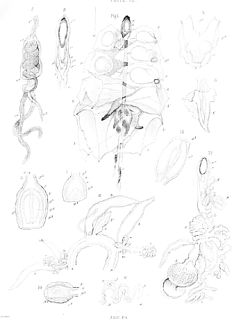
1. Agalma breve, complete: magnified. Taken on the east coast of Australia.
2. Its pneumatophore.
3. A fully formed and (figs. 4 and 5) young nectocalyces.
6. A young hydrophyllium.
7. The end of a tentacular branch.
8. Part of the cœnosarc, with hydrocysts and gonoblastidia.
9. 10. Young and fully formed gynophores.
11. An androphore.
12. A young Agalma .
|
THE
HUXLEY
FILE
|
| ||||||||||||||||||||||||||||||||||||||||||||||||||||||